

Lumbrokinase, a Fibrinolytic Enzyme, Prevents Intra-Abdominal Adhesion by Inhibiting the Migrative and Adhesive Activities of Fibroblast via Attenuation of the AP-1/ICAM-1 Signaling Pathway
- PMID: 36685669
- PMCID: PMC9851794
- DOI: 10.1155/2023/4050730
Lumbrokinase, a Fibrinolytic Enzyme, Prevents Intra-Abdominal Adhesion by Inhibiting the Migrative and Adhesive Activities of Fibroblast via Attenuation of the AP-1/ICAM-1 Signaling Pathway
Abstract
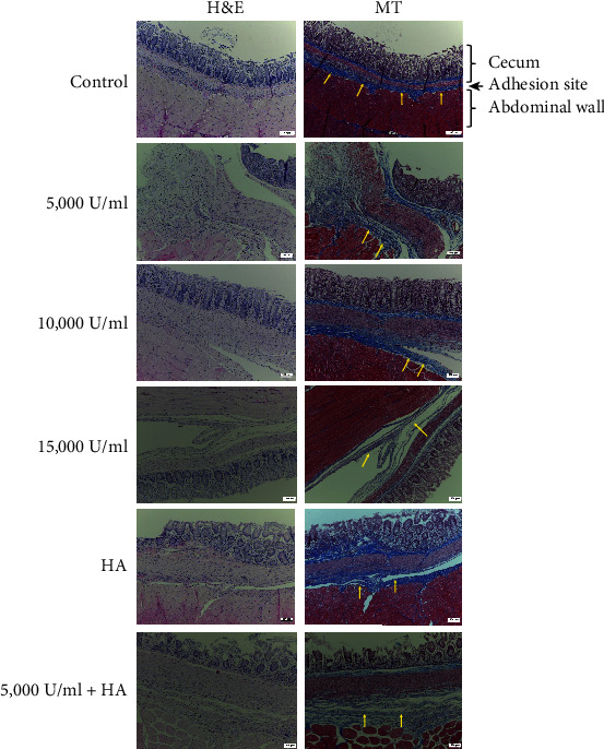
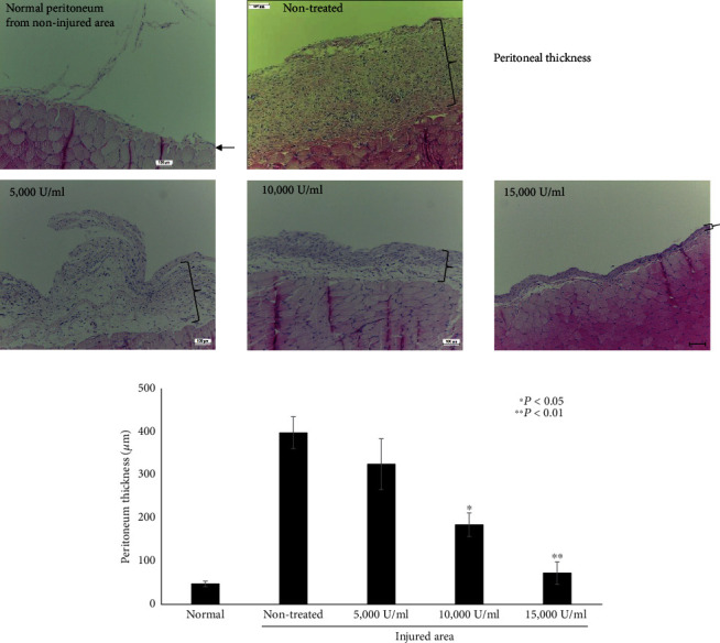
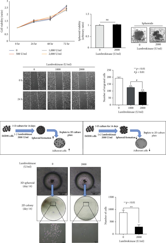
Intra-abdominal adhesion is a complication following abdominal surgery caused by the suppression of fibrinolytic activity and aggravated fibroblast invasion of the injured area, which may lead to chronic illnesses such as chronic pain, intestinal obstruction, and female infertility. This study hypothesized that lumbrokinase, a fibrinolytic enzyme extracted from the earthworm, supports the wound healing process. Therefore, we assessed the effect of lumbrokinase on intra-abdominal adhesion. Lumbrokinase treatment significantly decreased the severity and the area of intra-abdominal adhesion in vivo in a dose-dependent manner compared with the controls (untreated and hyaluronate-treated). Lumbrokinase-associated adverse effects were not observed. Immunohistochemical analysis of adhesion tissues revealed a loosened adhesive band between tissues, coupled with significantly decreased peritoneal thickening in the lumbrokinase-treated group versus the control group. Three-dimensional spheroid, MTT, and scratch wound migration assays using the IMR-90 human fibroblast cell line demonstrated that lumbrokinase significantly attenuated the migration and adhesive activity of fibroblasts without compromising cell proliferation. The luciferase assay and western blot analysis showed that lumbrokinase inhibited the AP-1/ICAM-1 cell adhesion signaling pathway. Therefore, lumbrokinase decreases intra-abdominal adhesion and peritoneal thickening by augmenting fibrinolytic action and inhibiting fibroblast migration and adhesive activity via attenuation of the AP-1/ICAM-1 signaling pathway. Lumbrokinase is thus a promising agent to prevent intra-abdominal adhesion.
Copyright © 2023 Que Thanh Thanh Nguyen et al.
Conflict of interest statement
The authors declare no conflicts of interest.
Figures

Similar articles
-
Intestinal Absorption of Fibrinolytic and Proteolytic Lumbrokinase Extracted from Earthworm, Eisenia andrei.Korean J Physiol Pharmacol. 2010 Apr;14(2):71-5. doi: 10.4196/kjpp.2010.14.2.71. Epub 2010 Apr 30. Korean J Physiol Pharmacol. 2010. PMID: 20473377 Free PMC article.
-
Antithrombogenicity of lumbrokinase-immobilized polyurethane.J Biomed Mater Res. 1994 Sep;28(9):1069-77. doi: 10.1002/jbm.820280912. J Biomed Mater Res. 1994. PMID: 7814434
-
Interleukin-1beta induces ICAM-1 expression enhancing leukocyte adhesion in human rheumatoid arthritis synovial fibroblasts: involvement of ERK, JNK, AP-1, and NF-kappaB.J Cell Physiol. 2010 Aug;224(2):516-26. doi: 10.1002/jcp.22153. J Cell Physiol. 2010. PMID: 20432452
-
Recombinant protein production of earthworm lumbrokinase for potential antithrombotic application.Evid Based Complement Alternat Med. 2013;2013:783971. doi: 10.1155/2013/783971. Epub 2013 Dec 12. Evid Based Complement Alternat Med. 2013. PMID: 24416067 Free PMC article. Review.
-
Oxidant stress regulation of IL-8 and ICAM-1 gene expression: differential activation and binding of the transcription factors AP-1 and NF-kappaB (Review).Int J Mol Med. 1999 Sep;4(3):223-30. doi: 10.3892/ijmm.4.3.223. Int J Mol Med. 1999. PMID: 10425270 Review.
Cited by
-
Natural serine proteases and their applications in combating amyloid formation.ADMET DMPK. 2024 Nov 16;12(6):797-820. doi: 10.5599/admet.2551. eCollection 2024. ADMET DMPK. 2024. PMID: 39713256 Free PMC article. Review.
-
Maggot Kinase and Natural Thrombolytic Proteins.ACS Omega. 2024 May 2;9(20):21768-21779. doi: 10.1021/acsomega.4c01663. eCollection 2024 May 21. ACS Omega. 2024. PMID: 38799322 Free PMC article. Review.
-
Promoted Skin Wound Healing by Tail-Amputated Eisenia foetida Proteins via the Ras/Raf/MEK/ERK Signaling Pathway.ACS Omega. 2023 Apr 5;8(15):13935-13943. doi: 10.1021/acsomega.3c00317. eCollection 2023 Apr 18. ACS Omega. 2023. PMID: 37091432 Free PMC article.
-
Leveraging Therapeutic Proteins and Peptides from Lumbricus Earthworms: Targeting SOCS2 E3 Ligase for Cardiovascular Therapy through Molecular Dynamics Simulations.Int J Mol Sci. 2024 Oct 8;25(19):10818. doi: 10.3390/ijms251910818. Int J Mol Sci. 2024. PMID: 39409145 Free PMC article.
-
Lumbrokinase Extracted from Earthworms Synergizes with Bevacizumab and Chemotherapeutics in Treating Non-Small Cell Lung Cancer by Targeted Inactivation of BPTF/VEGF and NF-κB/COX-2 Signaling.Biomolecules. 2024 Jun 23;14(7):741. doi: 10.3390/biom14070741. Biomolecules. 2024. PMID: 39062456 Free PMC article.
References
MeSH terms
Substances
LinkOut - more resources
Full Text Sources
