. 2023 Jan 4:13:1082709.
doi: 10.3389/fgene.2022.1082709. eCollection 2022.

Bioinformatic analysis and machine learning to identify the diagnostic biomarkers and immune infiltration in adenomyosis

Affiliations

Bioinformatic analysis and machine learning to identify the diagnostic biomarkers and immune infiltration in adenomyosis

Dan Liu et al. Front Genet. .

Abstract

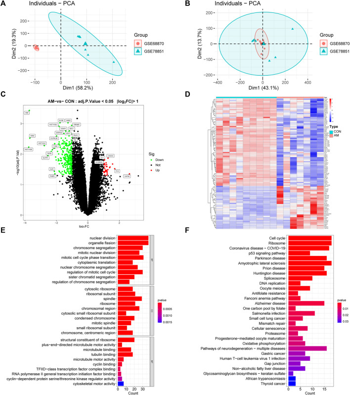
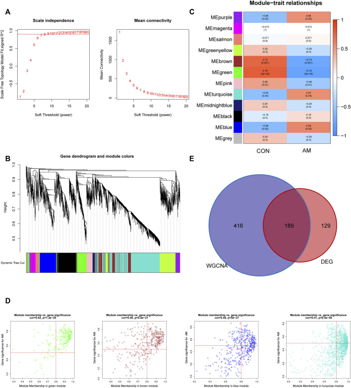
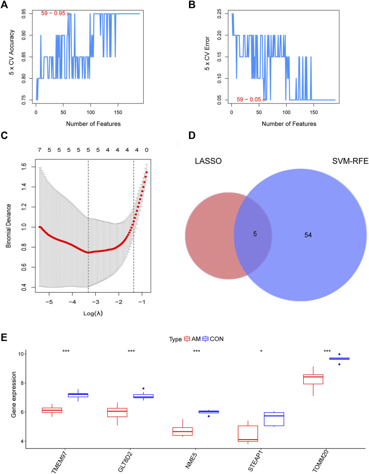
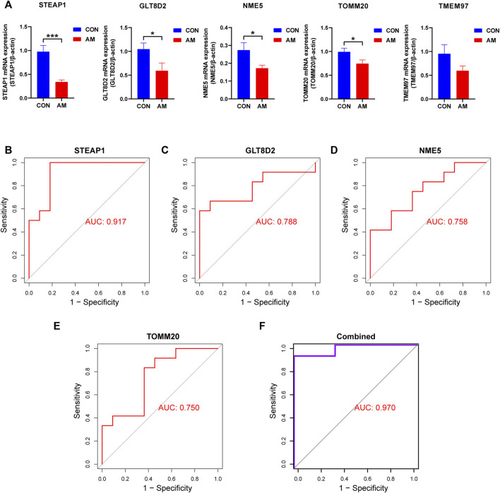
Background: Adenomyosis is a hormone-dependent benign gynecological disease characterized by the invasion of the endometrium into the myometrium. Women with adenomyosis can suffer from abnormal uterine bleeding, severe pelvic pain, and subfertility or infertility, which can interfere with their quality of life. However, effective diagnostic biomarkers for adenomyosis are currently lacking. The aim of this study is to explore the mechanism of adenomyosis by identifying biomarkers and potential therapeutic targets for adenomyosis and analyzing their correlation with immune infiltration in adenomyosis. Methods: Two datasets, GSE78851 and GSE68870, were downloaded and merged for differential expression analysis and functional enrichment analysis using R software. Weighted gene co-expression network analysis (WGCNA), the least absolute shrinkage and selection operator (LASSO), and support vector machine-recursive feature elimination (SVE-RFE) were combined to explore candidate genes. Quantitative reverse transcriptase PCR (qRT-PCR) was conducted to verify the biomarkers and receiver operating characteristic curve analysis was used to assess the diagnostic value of each biomarker. Single-sample Gene Set Enrichment Analysis (ssGSEA) and CIBERSORT were used to explore immune cell infiltration in adenomyosis and the correlation between diagnostic biomarkers and immune cells. Results: A total of 318 genes were differentially expressed. Through the analysis of differentially expressed genes and WGCNA, we obtained 189 adenomyosis-related genes. After utilizing the LASSO and SVM-RFE algorithms, four hub genes, namely, six-transmembrane epithelial antigen of the prostate-1 (STEAP1), translocase of outer mitochondrial membrane 20 (TOMM20), glycosyltransferase eight domain-containing 2 (GLT8D2), and NME/NM23 family member 5 (NME5) expressed in nucleoside-diphosphate kinase, were identified and verified by qRT-PCR. Immune infiltration analysis indicated that T helper 17 cells, CD56dim natural killer cells, monocytes, and memory B-cell may be associated with the occurrence of adenomyosis. There were significant correlations between the diagnostic biomarkers and immune cells. Conclusion: STEAP1, TOMM20, GLT8D2, and NME5 were identified as potential biomarkers and therapeutic targets for adenomyosis. Immune infiltration may contribute to the onset and progression of adenomyosis.

Keywords: WGCNA; adenomyosis; bioinformatics analysis; diagnostic markers; immune infiltration; machine learning.


Conflict of interest statement

The authors declare that the research was conducted in the absence of any commercial or financial relationships that could be construed as a potential conflict of interest.

Figures

FIGURE 1
FIGURE 1
Flowchart of this study. Microarray datasets analysis was conducted for endometrium samples from nine women with adenomyosis and seven healthy controls. DEGs: differentially expressed genes; GEO: Gene Expression Omnibus; GO: Gene Ontology; KEGG: Kyoto Encyclopedia of Genes and Genomes; LASSO: the least absolute shrinkage and selection operator; qRT-PCR: quantitative reverse transcriptase PCR; ROC: receiver operating characteristic; SVM-RFE: the support vector machine-recursive feature elimination; WGCNA: weighted gene co-expression network analysis.
FIGURE 2
FIGURE 2
Analysis of DEGs profile in endometrium between women with adenomyosis and controls. (A) The PCA plot of sample distribution from the two datasets before removing the batch effects. (B) The PCA plot of sample distribution from the two datasets after removing the batch effects. Different colors represent different datasets. (C) Volcano plot of DEGs. The red dots represent the up-regulated genes and the green dots represent the down-regulated genes in the adenomyosis group. (|log2 FC | ≥ 1; adjusted p-value <0.05). (D) Heatmap of DEGs; red indicates upregulated genes and blue indicates downregulated genes in the adenomyosis group. (E) GO enrichment analysis of DEGs. The top 10 BP, MF, and CC terms of DEGs. (F) The top 30 KEGG pathway enrichment analysis of DEGs. AM: adenomyosis group; BP: biological process; CC: cellular component; CON: control group; DEGs: differentially expressed genes; FC: fold change; GO: Gene Ontology; KEGG: Kyoto Encyclopedia of Genes and Genomes; MF: molecular function; PCA: principal component analysis.
FIGURE 3
FIGURE 3
Construction of gene co-expression networks associated with adenomyosis through WGCNA. (A) Determination of the soft-thresholding power (β). The analysis of the scale-free fit index for different soft-thresholding powers is shown in the left panel, and the mean connectivity for different soft-thresholding powers is shown in the right panel. (B) The cluster dendrogram of genes based on the dissimilarity of TOM. (C) The heatmap of correlation between genes in different modules and the clinical traits. (D) The correlation between GS for AM and MM in four modules (blue, purple, green and brown). One plot represents one gene. The criteria were set as |MM| > 0.8 and |GS| > 0.5. (E) Venn diagram of the hub genes obtained by intersecting DEGs and the adenomyosis-related key module genes identified by WGCNA. DEGs: differentially expressed genes; GS: gene significance; MM: module membership; TOM: topological overlap measure; WGCNA: weighted gene co-expression network analysis.
FIGURE 4
FIGURE 4
Identification of candidate diagnostic biomarkers by a comprehensive strategy. (A,B) Optimal genes identified using the SVM-RFE algorithm. (C) Significant prognostic variables screened using the LASSO regression. (D) Venn diagram of candidate diagnostic biomarkers screened using LASSO and SVM-RFE. (E) The expression of TMEM97, GLT8D2, NME5, STEAP1, and TOMM20 in microarray datasets (*p < 0.05, **p < 0.01, ***p < 0.001, ****p < 0.0001). GLT8D2: glycosyltransferase eight domain-containing two; LASSO: the least absolute shrinkage and selection operator; NDPK: nucleoside-diphosphate kinase; NME5: NME/NM23 family member five expressed in nucleoside-diphosphate kinase; STEAP1: six-transmembrane epithelial antigen of the prostate-1; SVM-RFE: support vector machine-recursive feature elimination; TOMM20: translocase of outer mitochondrial membrane 20; TMEM97: transmembrane protein 97.
FIGURE 5
FIGURE 5
Validation of hub genes using qRT-PCR. (A) Validation of the expression of candidate diagnostic biomarkers using qRT-PCR. Four diagnostic biomarkers, namely, STEAP1, GLT8D2, NME5, and TOMM20, were downregulated significantly in the endometrium of women with adenomyosis compared with the control group. The downregulation of TMEM97 did not show statistical significance. (B–E) The ROC curve analysis and calculation of the AUC of STEAP1, GLT8D2, NME5, and TOMM20 in the clinical samples. (F) The ROC curve to verify the diagnostic efficacy of the combined four diagnostic markers using logistic regression analysis (*p < 0.05, **p < 0.01, ***p < 0.001, ****p < 0.0001). AUC: area under the curve; GLT8D2: glycosyltransferase eight domain-containing two; NDPK: nucleoside-diphosphate kinase; NME5: NME/NM23 family member five expressed in nucleoside-diphosphate kinase; STEAP1: six-transmembrane epithelial antigen of the prostate-1; SVM-RFE: support vector machine-recursive feature elimination; TOMM20: translocase of outer mitochondrial membrane 20; TMEM97: transmembrane protein 97; qRT-PCR: quantitative reverse transcriptase PCR; ROC: receiver operating characteristic.
FIGURE 6
FIGURE 6
Immune cell infiltration analysis using ssGSEA. (A) Heatmap of the distribution of 28 immune cells in the adenomyosis and control group. (B) The violin plot of the different distribution of 28 immune cells between the adenomyosis and control groups (*p < 0.05, **p < 0.01, ***p < 0.001, ****p < 0.0001). SsGSEA: Single-sample Gene Set Enrichment Analysis.
FIGURE 7
FIGURE 7
Correlation between diagnostic biomarkers and infiltrating immune cells using CIBERSORT. (A) The bar plot of proportion of 22 immune cells in the endometrium of women with adenomyosis and control group analyzed using CIBERSORT. (B–D) The correlation between STEAP1, GLT8D2, TOMM20 and infiltrating immune cells. GLT8D2: glycosyltransferase eight domain-containing 2, STEAP1: six-transmembrane epithelial antigen of the prostate-1, TOMM20: translocase of outer mitochondrial membrane 20.

Similar articles

Cited by

References

    1. An M., Li D., Yuan M., Li Q., Zhang L., Wang G. (2017). Interaction of macrophages and endometrial cells induces epithelial-mesenchymal transition-like processes in adenomyosis. Biol. Reprod. 96 (1), 46–57. 10.1095/biolreprod.116.144071 - DOI - PubMed
    1. Anderegg L., Im Hof Gut M., Hetzel U., Howerth E. W., Leuthard F., Kyostila K., et al. (2019). NME5 frameshift variant in Alaskan Malamutes with primary ciliary dyskinesia. PLoS Genet. 15 (9), e1008378. 10.1371/journal.pgen.1008378 - DOI - PMC - PubMed
    1. Bai Z., Xu L., Dai Y., Yuan Q., Zhou Z. (2021). ECM2 and GLT8D2 in human pulmonary artery hypertension: Fruits from weighted gene co-expression network analysis. J. Thorac. Dis. 13 (4), 2242–2254. 10.21037/jtd-20-3069 - DOI - PMC - PubMed
    1. Bakhtiarizadeh M. R., Hosseinpour B., Shahhoseini M., Korte A., Gifani P. (2018). Weighted gene Co-expression network analysis of endometriosis and identification of functional modules associated with its main hallmarks. Front. Genet. 9, 453. 10.3389/fgene.2018.00453 - DOI - PMC - PubMed
    1. Benagiano G., Brosens I. (2006). History of adenomyosis. Best. Pract. Res. Clin. Obstet. Gynaecol. 20 (4), 449–463. 10.1016/j.bpobgyn.2006.01.007 - DOI - PubMed