

Upper gastrointestinal bleeding as an unusual manifestation of localized Ménétrier's disease with an underlying lipoma: A case report
- PMID: 36686066
- PMCID: PMC9846829
- DOI: 10.4253/wjge.v15.i1.10
Upper gastrointestinal bleeding as an unusual manifestation of localized Ménétrier's disease with an underlying lipoma: A case report
Abstract
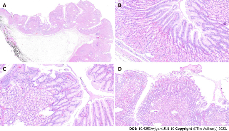
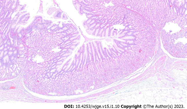
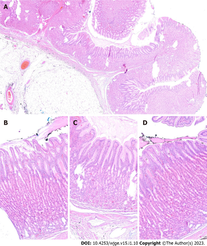
Background: Ménétrier's disease is a rare condition characterized by enlarged gastric folds, usually located in the whole body and fundus of the stomach. This report presents an unusual case of localized Ménétrier's disease elevated by a submucosal lipoma and thus looking like a polypoid mass and causing an episode of upper gastrointestinal bleeding. The mass was successfully removed with endoscopic submucosal dissection.
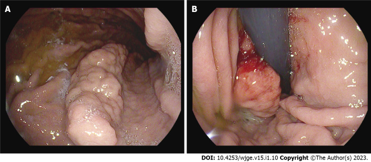
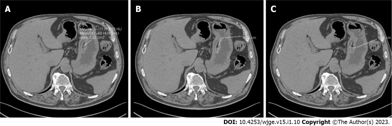
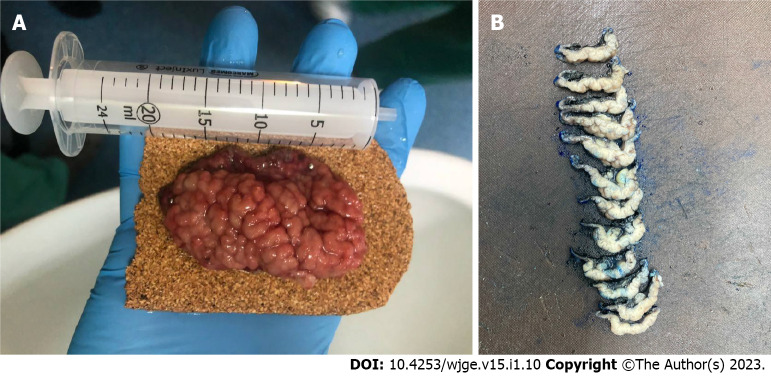
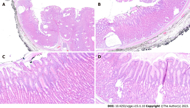
Case summary: Esophagogastroduodenoscopy was performed on a 76-year-old male patient after an episode of upper gastrointestinal bleeding, manifesting as fatigue and melena. A large polypoid mass (4 cm × 1 cm) with enlarged mucosal folds was found in the body of the stomach, between the lesser curvature and posterior wall. A small ulcer at the distal end of the mass was identified as the source of the bleeding. Biopsy was negative for neoplasia. Computed tomography showed a submucosal lesion beneath the affected mucosa, most likely a lipoma. The mass was removed en bloc with tunneling endoscopic submucosal dissection. Final pathology determined that the mass included Ménétrier's disease and a submucosal lipoma. The patient was scheduled for follow-up esophagogastroduodenoscopy.
Conclusion: Localized Ménétrier's disease can coexist with a submucosal lipoma creating a polypoid mass with risk of bleeding.
Keywords: Case report; Endoscopic submucosal dissection; Gastrointestinal hemorrhage; Ménétrier’s disease; Submucosal lipoma; Submucosal tunneling endoscopic resection.
©The Author(s) 2023. Published by Baishideng Publishing Group Inc. All rights reserved.
Conflict of interest statement
Conflict-of-interest statement: The authors declare that they have no conflict of interest.
Figures

References
-
- Lambrecht NW. Ménétrier's disease of the stomach: a clinical challenge. Curr Gastroenterol Rep. 2011;13:513–517. - PubMed
-
- Almazar AE, Penfield JD, Saito YA, Talley NJ. Survival Times of Patients With Menetrier's Disease and Risk of Gastric Cancer. Clin Gastroenterol Hepatol. 2021;19:707–712. - PubMed
-
- Dempsey PJ, Goldenring JR, Soroka CJ, Modlin IM, McClure RW, Lind CD, Ahlquist DA, Pittelkow MR, Lee DC, Sandgren EP. Possible role of transforming growth factor alpha in the pathogenesis of Ménétrier's disease: supportive evidence form humans and transgenic mice. Gastroenterology. 1992;103:1950–1963. - PubMed
Publication types
LinkOut - more resources
Full Text Sources
Research Materials
