. 2023 Jan 4:10:1031183.
doi: 10.3389/fbioe.2022.1031183. eCollection 2022.

Versatile electrical stimulator for cardiac tissue engineering-Investigation of charge-balanced monophasic and biphasic electrical stimulations

Affiliations

Versatile electrical stimulator for cardiac tissue engineering-Investigation of charge-balanced monophasic and biphasic electrical stimulations

Stefano Gabetti et al. Front Bioeng Biotechnol. .

Abstract

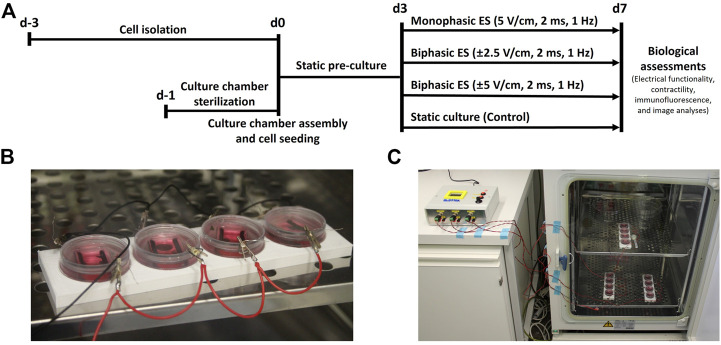
The application of biomimetic physical stimuli replicating the in vivo dynamic microenvironment is crucial for the in vitro development of functional cardiac tissues. In particular, pulsed electrical stimulation (ES) has been shown to improve the functional properties of in vitro cultured cardiomyocytes. However, commercially available electrical stimulators are expensive and cumbersome devices while customized solutions often allow limited parameter tunability, constraining the investigation of different ES protocols. The goal of this study was to develop a versatile compact electrical stimulator (ELETTRA) for biomimetic cardiac tissue engineering approaches, designed for delivering controlled parallelizable ES at a competitive cost. ELETTRA is based on an open-source micro-controller running custom software and is combinable with different cell/tissue culture set-ups, allowing simultaneously testing different ES patterns on multiple samples. In particular, customized culture chambers were appositely designed and manufactured for investigating the influence of monophasic and biphasic pulsed ES on cardiac cell monolayers. Finite element analysis was performed for characterizing the spatial distributions of the electrical field and the current density within the culture chamber. Performance tests confirmed the accuracy, compliance, and reliability of the ES parameters delivered by ELETTRA. Biological tests were performed on neonatal rat cardiac cells, electrically stimulated for 4 days, by comparing, for the first time, the monophasic waveform (electric field = 5 V/cm) to biphasic waveforms by matching either the absolute value of the electric field variation (biphasic ES at ±2.5 V/cm) or the total delivered charge (biphasic ES at ±5 V/cm). Findings suggested that monophasic ES at 5 V/cm and, particularly, charge-balanced biphasic ES at ±5 V/cm were effective in enhancing electrical functionality of stimulated cardiac cells and in promoting synchronous contraction.

Keywords: biomimetic approach; biphasic waveform; cardiac tissue engineering; charge balance; electrical stimulation; in vitro models; monophasic waveform; parallel investigation.


Conflict of interest statement

The authors declare that the research was conducted in the absence of any commercial or financial relationships that could be construed as a potential conflict of interest.

Figures

FIGURE 1
FIGURE 1
ELETTRA and culture chamber. (A) Schematic drawing of ELETTRA showing the relations between its subsystems and components. (B) Picture of the ELETTRA electrical stimulator. (C) Picture of the assembled culture chamber.
FIGURE 2
FIGURE 2
Biological experiments. (A) Timeline of the performed culture protocols. (B) Picture of four culture chambers connected in parallel during the experiments. (C) Picture of the whole setup during the biological experiments: each output of ELETTRA is connected to a set of four culture chambers placed inside the incubator.
FIGURE 3
FIGURE 3
Measurements of pulse train, voltage waveform and resulting current waveform on a single culture chamber for monophasic ES (A,B and C) and biphasic ES (D,E and F).
FIGURE 4
FIGURE 4
Comparison of measured currents and lumped parameter model results for multiple chambers connected in parallel to ELETTRA. (A) Current waveforms for 1, 3, and six chambers connected. (B) Peak currents for 1 to six chambers connected.
FIGURE 5
FIGURE 5
Distributions of the electric field and the current density within the culture chamber. (A) Contour plot of electric field magnitude at three planes perpendicular to the electrodes’ main axes, located at x = −10 mm, x = 0 mm, x = 10 mm. (B and C) Electric field magnitude on the central plane of the chamber along a line at the height of the electrodes centers and at the bottom of the chamber, respectively. (D) Vector diagram of the current density over the central cross section of the culture chamber.
FIGURE 6
FIGURE 6
Electrical functionality. (A) Excitation threshold (ET) and (B) Maximum capture rate (MCR) of CMs for the different culture conditions: Control (no stimulation; n. of replicates = 7); Monophasic ES (5V/cm, 1 Hz, 2 ms; n. of replicates = 8); Biphasic ES (±2.5V/cm, 1 Hz, 2 ms; n. of replicates = 6); Biphasic ES (±5V/cm, 1 Hz, 2 ms; n. of replicates = 7). Results from two independent experiments. Asterisks (*) denote statistically significant difference (*p < 0.05, **p < 0.01, ***p < 0.001).
FIGURE 7
FIGURE 7
Cardiomyocyte contractility. (A) Peak amplitude (PA) and (B) contraction time delay (CTD) of CMs for the different culture conditions: Control (no stimulation; n. of replicates = 3); Monophasic ES (5V/cm, 1 Hz, 2 ms; n. of replicates = 4); Biphasic ES (±2.5V/cm, 1 Hz, 2 ms; n. of replicates = 3); Biphasic ES (±5V/cm, 1 Hz, 2 ms; n. of replicates = 3). Asterisks (*) denote statistically significant differences (*p < 0.05, **p < 0.01, ***p < 0.001, ****p < 0.0001).
FIGURE 8
FIGURE 8
Immunofluorescence images of neonatal rat CMs for the different culture conditions. The images show the Connexin-43 (cyan) with the nuclei in blue (DAPI), Sarcomeric α-actinin (red) with the nuclei in blue (DAPI) in separated images and the merged signals for each experimental group. Scale bar = 50 µm.
FIGURE 9
FIGURE 9
Image-based quantification. (A) Percentage of area positive for Connexin-43, (B) percentage of area positive for Sarcomeric α-actinin, and (C) percentage of organized cardiomyocytes for the different culture conditions: Control (no stimulation; n. of replicates = 4); Monophasic ES (5V/cm, 1 Hz, 2 ms; n. of replicates = 4); Biphasic ES (±2.5V/cm, 1 Hz, 2 ms; n. of replicates = 4); Biphasic ES (±5V/cm, 1 Hz, 2 ms; n. of replicates = 4). Results from two independent experiments. Asterisks (*) denote statistically significant differences (*p < 0.05, **p < 0.01, ***p < 0.001, ****p < 0.0001).

Similar articles

Cited by

References

    1. Baumgartner S., Halbach M., Krausgrill B., Maass M., Srinivasan S. P., Sahito R. G. A., et al. (2015). Electrophysiological and morphological maturation of murine fetal cardiomyocytes during electrical stimulation in vitro . J. Cardiovasc. Pharmacol. Ther. 20, 104–112. 10.1177/1074248414536273 - DOI - PubMed
    1. Béland J., Duverger J. E., Petitjean E., Maguy A., Ledoux J., Comtois P. (2020). Development of an open hardware bioreactor for optimized cardiac cell culture integrating programmable mechanical and electrical stimulations. AIP Adv. 10, 035133. 10.1063/1.5144922 - DOI
    1. Birla R. k., Huang Y. c., Dennis R. g. (2007). Development of a novel bioreactor for the mechanical loading of tissue-engineered heart muscle. Tissue Eng. 13, 2239–2248. 10.1089/ten.2006.0359 - DOI - PubMed
    1. Buckberg G. D., Nanda N. C., Nguyen C., Kocica M. J. (2018). What is the heart? Anatomy, function, pathophysiology, and misconceptions. J. Cardiovasc. Dev. Dis. 5, 33. 10.3390/jcdd5020033 - DOI - PMC - PubMed
    1. Carlos-Oliveira M., Lozano-Juan F., Occhetta P., Visone R., Rasponi M. (2021). Current strategies of mechanical stimulation for maturation of cardiac microtissues. Biophys. Rev. 13, 717–727. 10.1007/s12551-021-00841-6 - DOI - PMC - PubMed

LinkOut - more resources