. 2023 Jan 5:10:1092543.
doi: 10.3389/fbioe.2022.1092543. eCollection 2022.

Gelatin-grafted tubular asymmetric scaffolds promote ureteral regeneration via activation of the integrin/Erk signaling pathway

Affiliations

Gelatin-grafted tubular asymmetric scaffolds promote ureteral regeneration via activation of the integrin/Erk signaling pathway

Baiyang Song et al. Front Bioeng Biotechnol. .

Abstract

Introduction: The repair of a diseased ureter is an urgent clinical issue that needs to be solved. A tissue-engineered scaffold for ureteral replacement is currently insufficient due to its incompetent bioactivity, especially in long-segment abnormalities. The primary reason is the failure of urothelialization on scaffolds. Methods: In this work, we investigated the ability of gelatin-grafted tubular scaffold in ureteral repairment and its related biological mechanism. We designed various porous asymmetric poly (L-lactic acid) (PLLA)/poly (L-lactide-co-e-caprolactone) (PLCL) tubes with a thermally induced phase separation (TIPS) method via a change in the ratio of solvents (named PP). To regulate the phenotype of urothelial cells and ureteral reconstruction, gelatin was grafted onto the tubular scaffold using ammonolysis and glutaraldehyde crosslinking (named PP-gel). The in vitro and in vivo experiments were performed to test the biological function and the mechanism of the scaffolds. Results and Discussion: The hydrophilicity of the scaffold significantly increased after gelatin grafting, which promoted the adhesion and proliferation of urothelial cells. Through subcutaneous implantation in rats, PP-gel scaffolds demonstrated good biocompatibility. The in vivo replacement showed that PP-gel could improve urothelium regeneration and maintain renal function after the ureter was replaced with an ∼4 cm-long PP-gel tube using New Zealand rabbits as the experimental animals. The related biologic mechanism of ureteral reconstruction was detected in detail. The gelatin-grafted scaffold upgraded the integrin α6/β4 on the urothelial cell membrane, which phosphorylates the focal adhesion kinase (FAK) and enhances urothelialization via the MAPK/Erk signaling pathway. Conclusion: All these results confirmed that the PP46-gel scaffold is a promising candidate for the constitution of an engineered ureter and to repair long-segment ureteral defects.

Keywords: MAPK/ERK pathway; gelatin; tissue engineering; ureter; urothelialization.


Conflict of interest statement

The authors declare that the research was conducted in the absence of any commercial or financial relationships that could be construed as a potential conflict of interest.

Figures

FIGURE 1
FIGURE 1
Procedure of PP-gel scaffold fabrication and the biomechanism of urothelialization. (A) Schematic illustration of the preparation of PLLA/PLCL scaffolds with grafted gelatin. (B) Biological mechanism of the PP-gel scaffold promoting urothelialization.
FIGURE 2
FIGURE 2
Characteristics of the PP46 and PP46-gel scaffold. (A) Pore diameter, wall thickness, and porosity as a function of the ratio of PLLA:PLCL. (B) Characteristics of the PP46 and PP46-gel. The morphology of scaffold PP46 and PP46-gel observed under SEM, cross section (a1-a2), and inner surface (b1-b2); (C, D) pore diameter and porosity of PP46-gel with scaffold PP46 as the reference (e1-e2). The EDS analysis of the inner surfaces of the PP46 and PP46-gel. (C) Contact angle alteration before and after gelatin grafting. (D) Quantitative analysis of the contact angles. (E) Stress–strain curves at a deformation rate of 10 mm/min. (F) Suture strength analysis. Data are presented as mean ± SD (**p < 0.01; ns: p > 0.05).
FIGURE 3
FIGURE 3
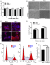
Proliferation of HUCs on the scaffold PP46 and PP46-gel. (A) Absorbance at a wavelength of 450 nm divided by the number of cells cultivated on the corresponding scaffolds. The cell number was measured with a hemocytometer. (B) Distribution of HUCs on the scaffolds observed under SEM. (C) Morphology of HUCs stained with F-actin (rhodamine-phalloidin, red) and nuclei (DAPI, blue). Scale bar, 20 μm. (D) Quantification of EGF secreted by cells on scaffolds PP46 or PP46-gel cultivated for 3 and 7 days, respectively. The value was divided by the number of cells cultivated on the corresponding scaffolds. (E) Cell cycle investigation tested using a flow cytometer. (F) Cell proliferation index (%) calculated from E. All data are presented as mean ± SD (*p < 0.05; ns: p > 0.05).
FIGURE 4
FIGURE 4
Histological analysis. (A) Scaffolds were implanted subcutaneously in SD rats for 4 and 8 weeks. Black arrows indicate CD68+ cells. (B, C) Quantitative analysis of the CD68+ ratio from IHC analysis. Scale bar, 400 μm (black) or 100 μm (white). Data are presented as mean ± SD (ns: p > 0.05; *p < 0.05).
FIGURE 5
FIGURE 5
Results of the in vivo tests. (A) Gross and tomography of PP46 and PP46-gel with the autograft as the positive control. Yellow arrows denote ureteral obstruction. Dotted circles are hydronephrosis images in CT. (B) Degree of hydronephrosis evaluated from CT cross-sectional scanning postoperatively at 12 weeks. (C, D) Assessments of the Cr and BUN content after material implantation. (E) Histological analysis. L, lumen. The triangles indicate the location where scaffolds were implanted. Black arrows indicate AE1/AE3+ cells. Scale bar, 100 μm. Data are presented as mean ± SD (*p < 0.05; **p < 0.01).
FIGURE 6
FIGURE 6
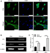
Phenotypic expression of HUCs on scaffolds PP46 and PP46-gel. (A) Epithelial phenotype with anti-cytokeratin AE1/AE3 as the primary antibody (green). The nuclei were stained with DAPI and displayed as blue. Cells were cultured for 7 days. Scale bar, 50 μm. (B) Analysis of UP3 and ZO-1 mRNA tested by RT-PCR. (C) Quantitative analysis of UP3 and ZO-1. Data are presented as mean ± SD (**p < 0.01).
FIGURE 7
FIGURE 7
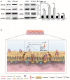
Studies of biological mechanisms. (A) Protein levels of ITGA6, ITGB4, FAK, p-FAK (Tyr 397), Ras, ERK, and p-ERK detected by Western blotting with GAPDH as the control. (B) Statistical analysis of ITGA6, ITGB4, p-FAK, Ras, and p-ERK. Cells were cultured for 7 days. (C) Schematic diagram regarding the molecular mechanism of the PP46-gel scaffold acting on the urothelial cells. Data are presented as mean ± SD (*p < 0.05; **p < 0.01).

Similar articles

Cited by

References

    1. Al-Maawi S., Rother S., Halfter N., Fiebig K. M., Moritz J., Moeller S., et al. (2022). Covalent linkage of sulfated hyaluronan to the collagen scaffold Mucograft® enhances scaffold stability and reduces proinflammatory macrophage activation in vivo . Bioact. Mater 8, 420–434. 10.1016/j.bioactmat.2021.06.008 - DOI - PMC - PubMed
    1. Dalghi M. G., Montalbetti N., Carattino M. D., Apodaca G. (2020). The urothelium: Life in a liquid environment. Physiol. Rev. 100 (4), 1621–1705. 10.1152/physrev.00041.2019 - DOI - PMC - PubMed
    1. Dalton G. D., Carney S. T., Marshburn J. D., Norford D. C., Howlett A. C. (2020). CB1 cannabinoid receptors stimulate gβγ-GRK2-mediated FAK phosphorylation at tyrosine 925 to regulate ERK activation involving neuronal focal adhesions. Front. Cell Neurosci. 14, 176. 10.3389/fncel.2020.00176 - DOI - PMC - PubMed
    1. de Jonge P., Simaioforidis V., Geutjes P. J., Oosterwijk E., Feitz W. F. J. (2014). Recent advances in ureteral tissue engineering. Curr. Urol. Rep. 16 (1), 465. 10.1007/s11934-014-0465-7 - DOI - PMC - PubMed
    1. de Jonge P., Sloff M., Janke H. P., Versteegden L. R. M., Kortmann B. B. M., de Gier R. P. E., et al. (2018). Ureteral reconstruction in goats using tissue-engineered templates and subcutaneous preimplantation. Tissue Eng. Part A 24 (11-12), 863–872. 10.1089/ten.TEA.2017.0347 - DOI - PubMed