P2X7R Mediates the Synergistic Effect of ATP and MSU Crystals to Induce Acute Gouty Arthritis
- PMID: 36686377
- PMCID: PMC9851801
- DOI: 10.1155/2023/3317307
P2X7R Mediates the Synergistic Effect of ATP and MSU Crystals to Induce Acute Gouty Arthritis
Abstract
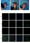
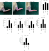
Activation of the nod-like receptor protein 3 (NLRP3) inflammasome by monosodium urate (MSU) crystals has been identified as the molecular basis for the acute inflammatory response in gouty arthritis. However, MSU crystals alone are not sufficient to induce acute gouty arthritis (AGA). Adenosine triphosphate (ATP) is an endogenous signaling molecule involved in the NLRP3 inflammasome activation. We aimed to explore the role of ATP in MSU crystal-induced AGA development. In peripheral blood mononuclear cell-derived macrophages obtained from gout patients, we observed a synergistic effect of ATP on MSU crystal-induced IL-1β release. Furthermore, in a rat model of spontaneous gout, we demonstrated that a synergistic effect of ATP and MSU crystals, but not MSU crystals alone, is essential for triggering AGA. Mechanistically, this synergistic effect is achieved through the purinergic receptor P2X7 (P2X7R). Blockade of P2X7R prevented AGA induction in rats after local injection of MSU crystals, and carrying the mutant hP2X7R gene contributed to the inhibition of NLRP3 inflammasome activation induced by costimulation of MSU crystals and ATP in vitro. Taken together, these results support the synergistic effect of ATP on MSU crystal-induced NLRP3 inflammasome activation facilitating inflammatory episodes in AGA. In this process, P2X7R plays a key regulatory role, suggesting targeting P2X7R to be an attractive therapeutic strategy for the treatment of AGA.
Copyright © 2023 Xiaoling Li et al.
Conflict of interest statement
The authors declare that the research was conducted in the absence of any commercial or financial relationships that could be construed as a potential conflict of interest.
Figures


Similar articles
-
Curcumin ameliorates monosodium urate-induced gouty arthritis through Nod-like receptor 3 inflammasome mediation via inhibiting nuclear factor-kappa B signaling.J Cell Biochem. 2019 Apr;120(4):6718-6728. doi: 10.1002/jcb.27969. Epub 2018 Dec 28. J Cell Biochem. 2019. PMID: 30592318
-
Gout-associated monosodium urate crystal-induced necrosis is independent of NLRP3 activity but can be suppressed by combined inhibitors for multiple signaling pathways.Acta Pharmacol Sin. 2022 May;43(5):1324-1336. doi: 10.1038/s41401-021-00749-7. Epub 2021 Aug 10. Acta Pharmacol Sin. 2022. PMID: 34376811 Free PMC article.
-
Doliroside A attenuates monosodium urate crystals-induced inflammation by targeting NLRP3 inflammasome.Eur J Pharmacol. 2014 Oct 5;740:321-8. doi: 10.1016/j.ejphar.2014.07.023. Epub 2014 Jul 24. Eur J Pharmacol. 2014. PMID: 25064339
-
P2X7R: a potential key regulator of acute gouty arthritis.Semin Arthritis Rheum. 2013 Dec;43(3):376-80. doi: 10.1016/j.semarthrit.2013.04.007. Epub 2013 Jun 17. Semin Arthritis Rheum. 2013. PMID: 23786870 Review.
-
Beneficial Properties of Phytochemicals on NLRP3 Inflammasome-Mediated Gout and Complication.J Agric Food Chem. 2018 Jan 31;66(4):765-772. doi: 10.1021/acs.jafc.7b05113. Epub 2018 Jan 17. J Agric Food Chem. 2018. PMID: 29293001 Review.
Cited by
-
[Avitinib suppresses NLRP3 inflammasome activation and ameliorates septic shock in mice].Nan Fang Yi Ke Da Xue Xue Bao. 2025 Aug 20;45(8):1697-1705. doi: 10.12122/j.issn.1673-4254.2025.08.14. Nan Fang Yi Ke Da Xue Xue Bao. 2025. PMID: 40916531 Free PMC article. Chinese.
-
The Progress of Immune Cells-induced Inflammatory Response in Gout.Curr Pharm Des. 2025;31(31):2465-2480. doi: 10.2174/0113816128369016250306050522. Curr Pharm Des. 2025. PMID: 40148294 Review.
-
Suppression of P2X7R by Local Treatment Alleviates Acute Gouty Inflammation.J Inflamm Res. 2023 Aug 22;16:3581-3591. doi: 10.2147/JIR.S421548. eCollection 2023. J Inflamm Res. 2023. PMID: 37636273 Free PMC article.
-
Gut-immunity-joint axis: a new therapeutic target for gouty arthritis.Front Pharmacol. 2024 Feb 23;15:1353615. doi: 10.3389/fphar.2024.1353615. eCollection 2024. Front Pharmacol. 2024. PMID: 38464719 Free PMC article. Review.
-
Animal Models for the Investigation of P2X7 Receptors.Int J Mol Sci. 2023 May 4;24(9):8225. doi: 10.3390/ijms24098225. Int J Mol Sci. 2023. PMID: 37175933 Free PMC article. Review.
References
MeSH terms
Substances
LinkOut - more resources
Full Text Sources
Medical
Miscellaneous

