. 2022 Dec 19;26(1):105838.
doi: 10.1016/j.isci.2022.105838. eCollection 2023 Jan 20.

3D blood-brain barrier-organoids as a model for Lyme neuroborreliosis highlighting genospecies dependent organotropism

Affiliations

3D blood-brain barrier-organoids as a model for Lyme neuroborreliosis highlighting genospecies dependent organotropism

Yvonne Adams et al. iScience. .

Abstract

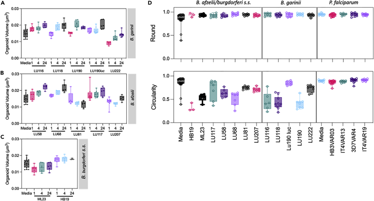
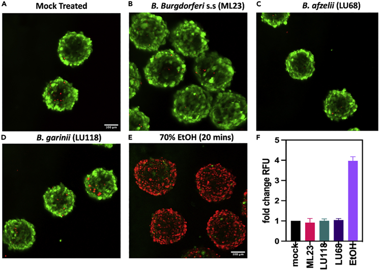
Lyme neuroborreliosis (LNB), a tick-borne infection caused by spirochetes within the Borrelia burgdorferi sensu lato (s.L.) complex, is among the most prevalent bacterial central nervous system (CNS) infections in Europe and the US. Here we have screened a panel of low-passage B. burgdorferi s.l. isolates using a novel, human-derived 3D blood-brain barrier (BBB)-organoid model. We show that human-derived BBB-organoids support the entry of Borrelia spirochetes, leading to swelling of the organoids and a loss of their structural integrity. The use of the BBB-organoid model highlights the organotropism between B. burgdorferi s.l. genospecies and their ability to cross the BBB contributing to CNS infection.

Keywords: Cell biology; Medical Microbiology; Neuroscience.


Conflict of interest statement

Disclosures: Outside of the present work: A.M.L. reports speaker’s honorarium/travel grants and advisory board activity from Gilead, GSK, and Pfizer. The other authors declare no competing interests exist. P.E.L. has been an external scientific expert to Valneva Austria GmbH and Pfizer Inc. and received speaker’s honorarium and travel grants.

Figures

None
Graphical abstract
Figure 1
Figure 1
Blood-brain barrier organoids support entry of Borrelia burgdorferi (A) 3D Z-projections of blood-brain barrier (BBB)-organoids (Magnification ×20) stained for Borrelia burgdorferi s.l. (CFSE, green), nuclei (DAPI, blue) and actin cytoskeleton (phalloidin, red). Scale bar 100 μm. (B left panel) Z-projection of BBB-organoid exposed to B. garinii LU116 for 4 h. White box and inset shows higher magnification image of single spirochete interacting with the surface of the organoid, lying perpendicular to the surface. Scale bar 100 μm. (B right panel) Orthogonal view from 54 μm within the organoid. White box shows invaded spirochete in Z, X, and Y axis. (C) Z-projections of BBB-organoids (magnification ×20) 24 h post-exposure to B. garinii LU116 showing the aggregation on the surface of each organoid. Each panel (A-D) shows a representative organoid from each group. ntotal organoids = 276.
Figure 2
Figure 2
Volumetric quantification of sub-surface Borrelia burgdorferi within blood-brain barrier-organoids (A) Graph plotting the total volume (μm3) of carboxyfluorescein succinimidyl ester (CFSE) signal within the organoids after 24 h exposure to B. garinii, B. afzelii, and B. burgdorferi s. s. (B) Maximum intensity Z-projection of CFSE signal from B. afzelii LU68 exposed organoid (magnification ×20). White asterisk denotes diffuse green stain indicative of phagocytosis. Scale bar 100 μm. ntotal organoids = 276.
Figure 3
Figure 3
Morphometric analysis of blood-brain barrier-organoids post-exposure with Borrelia burgdorferi The BBB organoids were co-incubated with ten different isolates of B. burgdorferi s. l., and two isolates of B. burgdorferi s. s. (Table 1), and red blood cells infected (IRBC) by four different P. falciparum lines. Box and whisker plots, showing min/max values (A–D) of BBB-organoid volume (μm3). Graphs (A–C) showed alterations post-Borrelia exposure. The gross morphology of organoids was measured by organoid roundness, i.e., general shape (D top panel), or the circularity i.e., organoid integrity. (D bottom panel). BBB-organoids were also exposed to P. falciparum lines (D both panels). The P. falciparum lines associated with cerebral (HB3VAR03, PFD1235w) has been shown to cross the BBB of organoids, while malaria parasites associated with uncomplicated (IT4VAR13, IT4VAR19) malaria did not. ntotal organoids = 276 (Borrelia), ntotal organoids = 61 (P. falciparum).
Figure 4
Figure 4
Internalization of Borrelia by blood-brain barrier-organoids Blood-brain barrier (BBB)-organoids co-incubated with B. burgdorferi s. l. isolates. (A) 3D Z-projection of organoids (magnification ×20) showing Carboxyfluorescein succinimidyl ester (CFSE stained) Borrelia in green, nuclei in blue (DAPI), and actin in red (phalloidin). The organoids were exposed to neurotropic (B. garinii LU118) or non-neurotrophic (B. afzelii LU207 and LU68) spirochetes. Composite images show overlays of CSFE, DAPI, and phalloidin stained images. (B) Orthogonal view of organoid exposed to B. garinii LU118.White filled arrows point toward a large aggregate of spirochetes extending from the surface and into the organoid. (C) Z-projection of BBB-organoid (magnification ×63) showing giant cell (white arrow) extending from the surface. Open arrows point to actin foci (phalloidin, red), whilst the asterisk denotes B. garinii LU116 spirochetes extending from surface into the cell (CFSE, green). Scale bar 100 μm. ntotal spheroids = 276 total.
Figure 5
Figure 5
Spirochete exposure leads to a loss of tight junctions Representative confocal images of organoids exposed to spirochetes for 24 h were fixed and then stained for the tight junction protein ZO-1 (green, AF488). (A) Z-projection of mock treated organoids showing bright ZO-1 stain around the periphery of the cells and diffuse within the cytosol. The dashed line from slices imaged at 90 μm depth (middle panel) represents the position analyzed to generate the intensity plots of ZO-1 stain (bottom panel). (B) Organoids exposed to vascular endothelial growth factor (VEGF), show disrupted tight junctions and diffuse stain, while those exposed to (C) B. burgdorferi s.s. ML23, (D) B. garinii LU118, or (E) B. afzelii LU68 show low intensity, diffuse stain (red, AF647) ntotal spheroids = 60 total. Scale bar = 100 μm.
Figure 6
Figure 6
Live/Dead analysis of organoids post-spirochete exposure (A–D) To determine the level of cell death post-spirochete exposure, organoids were treated with Live/Dead stain. Live cells in the organoids stain green, while dead cells stain red. Very few red stained are observed in the mock treated controls (A), or in the organoids exposed to (B) B. burgdorferi s.s. (B) ML23, (C) B. garinii LU118, or (D) B. afzelii LU68 spirochetes after 24 h co-incubation. The negative control organoids (E) were treated with 70% EtOH for 20 min and most cells were dead. (F) plot showing the fold change in cell death compared to mock treated controls (+/−SD), with very little change post-spirochete exposure. ntotal spheroids = 60 total. Scale bar = 100 μm.

References

    1. Kullberg B.J., Vrijmoeth H.D., van de Schoor F., Hovius J.W. Lyme borreliosis: diagnosis and management. BMJ. 2020;369:m1041. doi: 10.1136/bmj.m1041. - DOI - PubMed
    1. Berglund J., Eitrem R., Ornstein K., Lindberg A., Ringér A., Elmrud H., Carlsson M., Runehagen A., Svanborg C., Norrby R. An epidemiologic study of Lyme disease in southern Sweden. N. Engl. J. Med. 1995;333:1319–1327. doi: 10.1056/NEJM199511163332004. - DOI - PubMed
    1. Rupprecht T.A., Koedel U., Fingerle V., Pfister H.-W. The pathogenesis of Lyme neuroborreliosis: from infection to inflammation. Mol. Med. 2008;14:205–212. doi: 10.2119/2007-00091.Rupprecht. - DOI - PMC - PubMed
    1. Garcia-Monco J.C., Benach J.L. Lyme neuroborreliosis: clinical outcomes, controversy, pathogenesis, and polymicrobial infections. Ann. Neurol. 2019;85:21–31. doi: 10.1002/ana.25389. - DOI - PMC - PubMed
    1. Hansen K., Lebech A.-M. The Clinical and Epidemiological profile of Lyme Neuroborreliosis in Denmark 1985-1990: a prospective study of 187 patients with Borrelia burgdorferi specific intrathecal antibody production. Brain. 1992;115:399–423. doi: 10.1093/brain/115.2.399. - DOI - PubMed