. 2023 Jan 4:13:1028191.
doi: 10.3389/fendo.2022.1028191. eCollection 2022.

Growth hormone stimulates lipolysis in mice but not in adipose tissue or adipocyte culture

Affiliations

Growth hormone stimulates lipolysis in mice but not in adipose tissue or adipocyte culture

Lidan Zhao et al. Front Endocrinol (Lausanne). .

Abstract

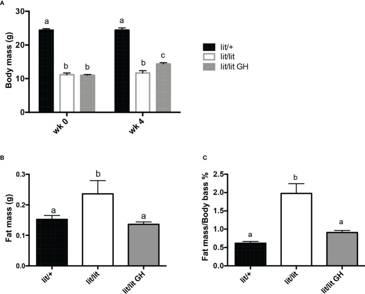
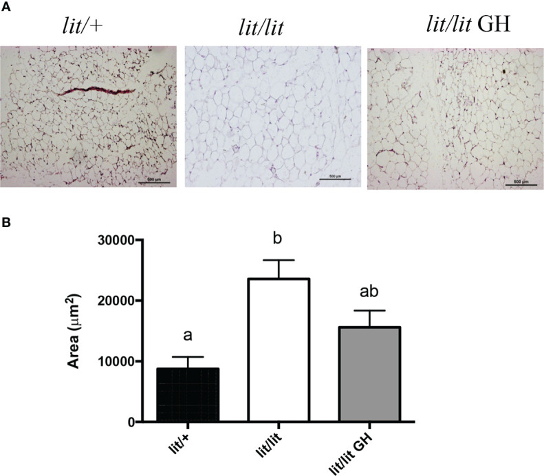
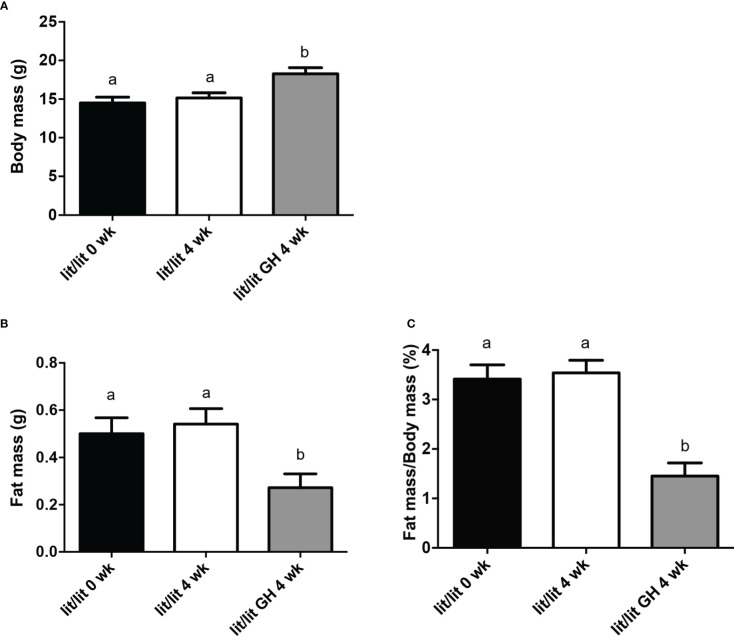
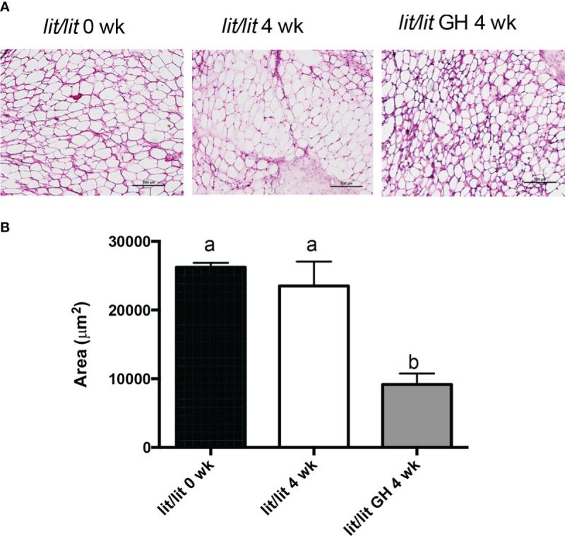
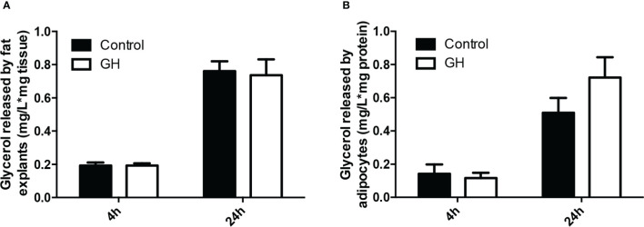
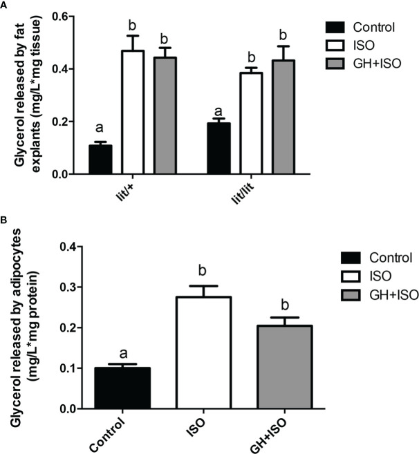
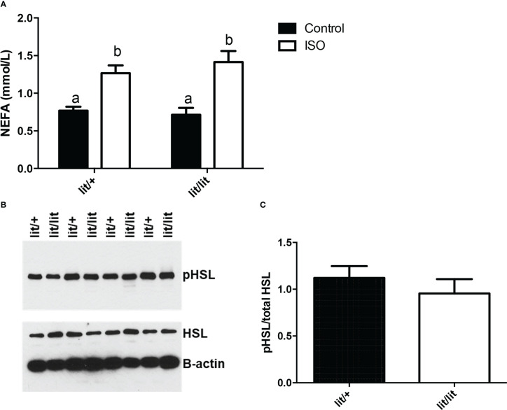
The inhibitory effect of growth hormone (GH) on adipose tissue growth and the stimulatory effect of GH on lipolysis are well known, but the mechanisms underlying these effects are not completely understood. In this study, we revisited the effects of GH on adipose tissue growth and lipolysis in the lit/lit mouse model. The lit/lit mice are GH deficient because of a mutation in the GH releasing hormone receptor gene. We found that the lit/lit mice had more subcutaneous fat and larger adipocytes than their heterozygous lit/+ littermates and that these differences were partially reversed by 4-week GH injection. We also found that GH injection to the lit/lit mice caused the mature adipose tissue and adipocytes to reduce in size. These results demonstrate that GH inhibits adipose tissue growth at least in part by stimulating lipolysis. To determine the mechanism by which GH stimulates lipolysis, we cultured adipose tissue explants and adipocytes derived from lit/lit mice with GH and/or isoproterenol, an agonist of the beta-adrenergic receptors. These experiments showed that whereas isoproterenol, expectedly, stimulated potent lipolysis, GH, surprisingly, had no effect on basal lipolysis or isoproterenol-induced lipolysis in adipose tissue explants or adipocytes. We also found that both isoproterenol-induced lipolysis and phosphorylation of hormone-sensitive lipase were not different between lit/lit and lit/+ mice. Taken together, these results support the conclusion that GH has lipolytic effect in mice but argue against the notion that GH stimulates lipolysis by directly acting on adipocytes or by enhancing β-adrenergic receptors-mediated lipolysis.

Keywords: adipocytes; adipose tissue; adrenergic; growth hormone; lipolysis.


Conflict of interest statement

The authors declare that the research was conducted in the absence of any commercial or financial relationships that could be construed as a potential conflict of interest.

Figures

Figure 1
Figure 1
Effects of GH deficiency and GH injection on body mass and fat mass of mice. Three groups of 9-week-old male lit/lit, lit/lit, and lit/+ mice from the same litters were injected with 2 μg/g body mass of recombinant bovine GH, vehicle control, and vehicle control, respectively, once a day for 4 weeks. Body mass and mass of inguinal subcutaneous fat pads were recorded at the beginning (wk 0) and end (wk 4) of the experiment. (A) Body mass. (B) Mass of inguinal subcutaneous fat pads. (C) Inguinal fat pad mass as percentage of body mass. Data are expressed as mean ± SEM (n = 5 mice). Means labeled with different letters are statistically different (P < 0.05).
Figure 2
Figure 2
Effects of GH deficiency and GH injection on average adipocyte size in mice. Three groups of 9-week-old male lit/lit, lit/lit, and lit/+ mice from the same litters were injected with GH, vehicle control, and vehicle control, respectively, once a day, for 4 weeks. Inguinal subcutaneous fat pads were collected at the end of the experiment for sectioning and staining. (A) Representative micrographs of fat sections. (B) Average adipocyte size. Data are expressed as means ± SEM (n = 5 mice). Means not sharing the same letter label are statistically different (P < 0.05).
Figure 3
Figure 3
Body mass and inguinal subcutaneous fat mass of lit/lit mice before and after GH injection. Body mass and mass of inguinal subcutaneous fat pads were recorded from three groups of mice: 17-week-old lit/lit mice (indicated as lit/lit 0 wk in the graphs), 21-week-old lit/lit mice injected with GH for 4 weeks (indicated as lit/lit GH 4 wk in the graphs), and 21-week-old lit/lit mice injected with control for 4 weeks (indicated as lit/lit 4 wk in the graphs). (A) Body mass. (B) Fat mass. (C) Fat mass as percentage of body mass. Data are expressed as means ± SEM (n = 4 mice). Means labeled with different letters are statistically different (P < 0.05).
Figure 4
Figure 4
Adipocyte size of lit/lit mice before and after GH injection. Inguinal subcutaneous fat pads from 17-week-old lit/lit mice, 21-week-old lit/lit mice injected with GH for 4 weeks, and 21-week-old lit/lit mice injected with control for 4 weeks, indicated as lit/lit 0 wk, lit/lit GH 4 wk, and lit/lit 4 wk, respectively, in the graphs, were sectioned and stained with hematoxylin and eosin. (A) Representative photos of adipose tissue sections. (B) Average area of adipocytes. Data are expressed as mean ± SEM (n = 4 mice). Means labeled with different letters are statistically different (P < 0.05).
Figure 5
Figure 5
Effect of GH on lipolysis in adipose tissue and adipocytes. (A) Effect of GH on lipolysis in adipose tissue explants. Inguinal subcutaneous fat explants from ad lib-fed lit/lit mice were cultured with vehicle control or 100 ng/ml GH for 4 h and 24 h, and glycerol concentration in the medium was measured. Concentration of glycerol in the medium was normalized by the mass of adipose tissue. (B) Effect of GH on lipolysis in adipocytes. The stromal vascular fraction cells were isolated from ad lib-fed lit/lit inguinal subcutaneous fat and differentiated into adipocytes in culture. Adipocytes were then treated with control or 100 ng/ml GH for 4 h and 24 h. Glycerol concentration was normalized by total protein content of adipocytes. Data are expressed as mean ± SEM (n = 5 mice). Both 4-h and 24-h GH treatments had no effect (P > 0.1) on glycerol release from either adipose tissue or adipocytes. * represents multiplication.
Figure 6
Figure 6
Effect of GH on isoproterenol-induced lipolysis in adipose tissue and adipocytes. (A) Effect of GH on isoproterenol-induced lipolysis in adipose tissue explants. Inguinal subcutaneous fat explants from ad lib-fed lit/+ mice and lit/lit littermates were cultured with 10 μM isoproterenol (ISO), 10 μM ISO and 100 ng/ml GH, or vehicle control for 4 h, and glycerol released into the medium was measured. (B) Effect of GH on isoproterenol-induced lipolysis in adipocytes. Adipocytes were differentiated from the stromal vascular fraction cells isolated from ad lib-fed lit/lit inguinal subcutaneous fat. Adipocytes were cultured with 10 μM ISO, 10 μM ISO and 100 ng/ml GH, or vehicle control for 4 h, and glycerol released into the medium was measured. Data are expressed as mean ± SEM (n = 5 mice). Means labeled with different letters are statistically different (P < 0.05). * represents multiplication.
Figure 7
Figure 7
Effect of GH deficiency on isoproterenol-induced lipolysis in mice. Ad lib-fed Lit/+ mice and lit/lit littermates were intraperitoneally injected with 10 μg/g body mass of isoproterenol (ISO). Blood samples were collected immediately before and 15 min after the injection, and inguinal subcutaneous fat pads were taken 15 min after the injection. (A) Serum concentrations of non-esterified fatty acids (NEFA). Means labeled with different letters are statistically different (P < 0.05, n = 4 mice). (B) Western blots of phosphorylated hormone-sensitive lipase (pHSL) and total HSL in adipose tissue. Beta-actin was measured as a loading control. (C) Density ratio of pHSL to total HSL. pHSL/HSL is not different between lit/+ and lit/lit mice.

Similar articles

Cited by

References

    1. Etherton TD, Louveau I, Sorensen MT, Chaudhuri S. Mechanisms by which somatotropin decreases adipose tissue growth. Am J Clin Nutr (1993) 58(2 Suppl):287S–95S. doi: 10.1093/ajcn/58.2.287S - DOI - PubMed
    1. Rosen T, Bosaeus I, Tolli J, Lindstedt G, Bengtsson BA. Increased body fat mass and decreased extracellular fluid volume in adults with growth hormone deficiency. Clin Endocrinol (Oxf) (1993) 38(1):63–71. doi: 10.1111/j.1365-2265.1993.tb00974.x - DOI - PubMed
    1. Berryman DE, List EO, Palmer AJ, Chung MY, Wright-Piekarski J, Lubbers E, et al. . Two-year body composition analyses of long-lived ghr null mice. J Gerontol A Biol Sci Med Sci (2010) 65(1):31–40. doi: 10.1093/gerona/glp175 - DOI - PMC - PubMed
    1. Bengtsson BA, Eden S, Lonn L, Kvist H, Stokland A, Lindstedt G, et al. . Treatment of adults with growth hormone (Gh) deficiency with recombinant human gh. J Clin Endocrinol Metab (1993) 76(2):309–17. doi: 10.1210/jcem.76.2.8432773 - DOI - PubMed
    1. Maison P, Griffin S, Nicoue-Beglah M, Haddad N, Balkau B, Chanson P. Impact of growth hormone (Gh) treatment on cardiovascular risk factors in gh-deficient adults: A metaanalysis of blinded, randomized, placebo-controlled trials. J Clin Endocrinol Metab (2004) 89(5):2192–9. doi: 10.1210/jc.2003-030840 - DOI - PubMed

LinkOut - more resources