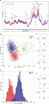
Raman spectroscopy: A prospective intraoperative visualization technique for gliomas
- PMID: 36686726
- PMCID: PMC9849680
- DOI: 10.3389/fonc.2022.1086643
Raman spectroscopy: A prospective intraoperative visualization technique for gliomas
Abstract
The infiltrative growth and malignant biological behavior of glioma make it one of the most challenging malignant tumors in the brain, and how to maximize the extent of resection (EOR) while minimizing the impact on normal brain tissue is the pursuit of neurosurgeons. The current intraoperative visualization assistance techniques applied in clinical practice suffer from low specificity, slow detection speed and low accuracy, while Raman spectroscopy (RS) is a novel spectroscopy technique gradually developed and applied to clinical practice in recent years, which has the advantages of being non-destructive, rapid and accurate at the same time, allowing excellent intraoperative identification of gliomas. In the present work, the latest research on Raman spectroscopy in glioma is summarized to explore the prospect of Raman spectroscopy in glioma surgery.
Keywords: EOR; Raman spectroscopy; SERS; SRH; glioma; intraoperative.
Copyright © 2023 Zhang, Yu, Li, Xu, Yang, Shan, Du, Yan and Chen.
Conflict of interest statement
The authors declare that the research was conducted in the absence of any commercial or financial relationships that could be construed as a potential conflict of interest.
Figures


Similar articles
-
Current Applications of Raman Spectroscopy in Intraoperative Neurosurgery.Biomedicines. 2024 Oct 16;12(10):2363. doi: 10.3390/biomedicines12102363. Biomedicines. 2024. PMID: 39457674 Free PMC article. Review.
-
Introduction of a standardized multimodality image protocol for navigation-guided surgery of suspected low-grade gliomas.Neurosurg Focus. 2015 Jan;38(1):E4. doi: 10.3171/2014.10.FOCUS14597. Neurosurg Focus. 2015. PMID: 25552284
-
A review of clinical use of surface-enhanced Raman scattering-based biosensing for glioma.Front Neurol. 2024 Apr 8;15:1287213. doi: 10.3389/fneur.2024.1287213. eCollection 2024. Front Neurol. 2024. PMID: 38651101 Free PMC article. Review.
-
Intraoperative perception and estimates on extent of resection during awake glioma surgery: overcoming the learning curve.J Neurosurg. 2018 May;128(5):1410-1418. doi: 10.3171/2017.1.JNS161811. Epub 2017 Jul 21. J Neurosurg. 2018. PMID: 28731401
-
Surgical benefits of combined awake craniotomy and intraoperative magnetic resonance imaging for gliomas associated with eloquent areas.J Neurosurg. 2017 Oct;127(4):790-797. doi: 10.3171/2016.9.JNS16152. Epub 2017 Jan 6. J Neurosurg. 2017. PMID: 28059650
Cited by
-
Raman Spectroscopy in the Diagnosis of Brain Gliomas: A Literature Review.Cureus. 2025 Feb 17;17(2):e79165. doi: 10.7759/cureus.79165. eCollection 2025 Feb. Cureus. 2025. PMID: 40109807 Free PMC article. Review.
-
Current Applications of Raman Spectroscopy in Intraoperative Neurosurgery.Biomedicines. 2024 Oct 16;12(10):2363. doi: 10.3390/biomedicines12102363. Biomedicines. 2024. PMID: 39457674 Free PMC article. Review.
-
Advancing Brain Research through Surface-Enhanced Raman Spectroscopy (SERS): Current Applications and Future Prospects.Biosensors (Basel). 2024 Jan 10;14(1):33. doi: 10.3390/bios14010033. Biosensors (Basel). 2024. PMID: 38248410 Free PMC article. Review.
-
Perspective: Raman spectroscopy for detection and management of diseases affecting the nervous system.Front Vet Sci. 2024 Oct 21;11:1468326. doi: 10.3389/fvets.2024.1468326. eCollection 2024. Front Vet Sci. 2024. PMID: 39497742 Free PMC article.
-
Development of a rapid and novel diagnostic technique for cardiac amyloidosis using Raman spectroscopy.Res Sq [Preprint]. 2025 Jun 25:rs.3.rs-6795517. doi: 10.21203/rs.3.rs-6795517/v1. Res Sq. 2025. PMID: 40678255 Free PMC article. Preprint.
References
-
- Coburger J, Merkel A, Scherer M, Schwartz F, Gessler F, Roder C, et al. . Low-grade glioma surgery in intraoperative magnetic resonance imaging: Results of a multicenter retrospective assessment of the German study group for intraoperative magnetic resonance imaging. Neurosurgery (2016) 78(6):775–86. doi: 10.1227/NEU.0000000000001081 - DOI - PubMed
-
- Fukui A, Muragaki Y, Saito T, Maruyama T, Nitta M, Ikuta S, et al. . Volumetric analysis using low-field intraoperative magnetic resonance imaging for 168 newly diagnosed supratentorial glioblastomas: Effects of extent of resection and residual tumor volume on survival and recurrence. World Neurosurg (2017) 98:73–80. doi: 10.1016/j.wneu.2016.10.109 - DOI - PubMed
Publication types
LinkOut - more resources
Full Text Sources
Miscellaneous

