Case report: Primary intracranial mucosa-associated lymphoid tissue lymphoma presenting as two primary tumors involving the cavernous sinus and extra-axial dura, respectively
- PMID: 36686768
- PMCID: PMC9846770
- DOI: 10.3389/fonc.2022.927086
Case report: Primary intracranial mucosa-associated lymphoid tissue lymphoma presenting as two primary tumors involving the cavernous sinus and extra-axial dura, respectively
Abstract
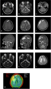
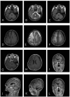
Primary intracranial mucosa-associated lymphoid tissue (MALT) lymphoma is a rare type of brain tumor, with only a few reported cases worldwide that mostly have only one lesion with conventional magnetic resonance imaging (MRI) findings. Here, we present a special case of intracranial MALT lymphoma with two mass lesions radiographically consistent with meningiomas on MRI before the operation. A 66-year-old woman was admitted to the hospital with intermittent right facial pain for 1 year, aggravated for the last month. Brain MRI showed two extracerebral solid masses with similar MR signal intensity. One mass was crescent-shaped beneath the skull, and the other was in the cavernous sinus area. Lesions showed isointensity on T1WI and T2WI and an intense homogeneous enhancement after contrast agent injection. Both lesions showed hyperintensity in amide proton transfer-weighted images. The two masses were all surgically resected. The postoperative pathology indicated extranodal marginal zone B-cell lymphoma of MALT. To improve awareness of intracranial MALT lymphoma in the differential diagnosis of extra-axial lesions among clinicians, we present this report and briefly summarize previously reported cases to describe the clinical, pathological, radiological, and treatment features.
Keywords: central nervous system; extranodal marginal zone lymphoma; magnetic resonance imaging; mucosa-associated lymphoid tissue; primary intracranial tumor; treatment.
Copyright © 2023 Tian, Pan, Gao, Li, Liu, Zou and Miao.
Conflict of interest statement
The authors declare that the research was conducted in the absence of any commercial or financial relationships that could be construed as a potential conflict of interest.
Figures


Similar articles
-
Extranodal marginal zone B-cell lymphoma of malt type involving the cavernous sinus.Leuk Lymphoma. 2001 Sep-Oct;42(5):1133-7. doi: 10.3109/10428190109097736. Leuk Lymphoma. 2001. PMID: 11697633 Review.
-
Primary Hepatic Extranodal Marginal Zone Lymphoma of Mucosa-Associated Lymphoid Tissue in a Patient with Chronic Hepatitis B Virus Infection: Case Report and Summary of the Literature.Medicina (Kaunas). 2021 Mar 18;57(3):280. doi: 10.3390/medicina57030280. Medicina (Kaunas). 2021. PMID: 33803501 Free PMC article.
-
Primary extranodal marginal zone mucosa-associated lymphoid tissue-type B-cell lymphoma involving the dura: A case report.Surg Neurol Int. 2024 Mar 29;15:113. doi: 10.25259/SNI_792_2023. eCollection 2024. Surg Neurol Int. 2024. PMID: 38628522 Free PMC article.
-
Primary marginal zone B-cell lymphoma of the cavernous sinus: a case report and review of the literature.BMC Med Imaging. 2021 Feb 12;21(1):25. doi: 10.1186/s12880-021-00556-w. BMC Med Imaging. 2021. PMID: 33579209 Free PMC article. Review.
-
Extranodal Marginal Zone B-Cell Lymphoma of Mucosa-Associated Tissue Type Involving the Dura.Cancer Res Treat. 2016 Apr;48(2):859-63. doi: 10.4143/crt.2014.334. Epub 2015 Jul 17. Cancer Res Treat. 2016. PMID: 26194368 Free PMC article.
Cited by
-
Intracranial Rosai‑Dorfman disease complicated by mucosa‑associated lymphoid tissue: A case report.Oncol Lett. 2023 Jul 31;26(3):402. doi: 10.3892/ol.2023.13987. eCollection 2023 Sep. Oncol Lett. 2023. PMID: 37600326 Free PMC article.
References
Publication types
LinkOut - more resources
Full Text Sources

