. 2023 Jan 4:12:1067312.
doi: 10.3389/fonc.2022.1067312. eCollection 2022.

Closure strategy for endoscopic pituitary surgery: Experience from 3015 patients

Affiliations

Closure strategy for endoscopic pituitary surgery: Experience from 3015 patients

Bertrand Baussart et al. Front Oncol. .

Abstract

Introduction: Effective strategies are required to ensure optimal management of the crucial closure step in endoscopic pituitary surgery. Many surgical techniques have been reported but no significant consensus has been defined.

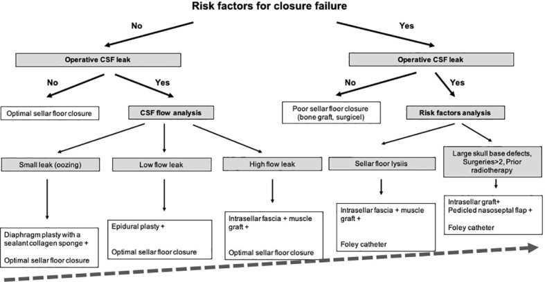
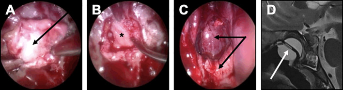
Methods: Between January 2006 and March 2022, 3015 adult patients with pituitary adenomas were operated on by a single expert neurosurgical team, using a mononostril endoscopic endonasal approach. Based of preoperative risk factors of and operative findings, a detailed closure strategy was used. Body mass index >40, sellar floor lysis, number of surgeries>2, large skull base destruction, prior radiotherapy were considered as preoperative risk factors for closure failure. All patients treated with an expanded endonasal approach were excluded.

Results: Patients were mostly women (F/M ratio: 1.4) with a median age of 50 (range: 18 -89). Intraoperative CSF leak requiring specific surgical management was observed in 319/3015 (10.6%) of patients. If intraoperative leak occurred, patients with predictive risk factors were managed using a Foley balloon catheter in case of sellar floor lysis or BMI>40 and a multilayer repair strategy with a vascularized nasoseptal flap in other cases. Postoperative CSF leak occurred in 29/3015 (1%) of patients, while meningitis occurred in 24/3015 (0.8%) of patients. In patients with intraoperative leak, closure management failed in 11/319 (3.4%) of cases.

Conclusion: Based on our significant 16-year experience, our surgical management is reliable and easy to follow. With a planned and stepwise strategy, the closure step can be optimized and tailored to each patient with a very low failure rate.

Keywords: closure; endoscopy; nasoseptal flap; pituitary surgery; skull base repair; strategy.


Conflict of interest statement

The authors declare that the research was conducted in the absence of any commercial or financial relationships that could be construed as a potential conflict of interest.

Figures

Figure 1
Figure 1
Preparation of the closure step during the endoscopic approach. This figure illustrates the concept that closure should be anticipated and planned for at each surgical step. (A) Sphenoid step. The main classic landmarks are identified: sella turcica (ST), tuberculum sellae (TuS), clivus (Cl), cavernous sinus (CS). (B) Sellar step. After opening the sellar floor, the dura has been carefully exposed and respected. The optimal rectangle shaped-dural opening is provided (dotted black rectangle). Note that the bone opening is oversized compared to the dural opening, in order to preserve the epidural space. (C) Sellar step after tumor resection. Note that the epidural space has been respected between the sellar floor and the dura mater (black arrow) for optimal closure.
Figure 2
Figure 2
Closure strategy for pituitary surgery. Decision strategy.
Figure 3
Figure 3
Closure strategy in patients with no CSF leak and no risk factor. For these patients, an optimal sellar floor closure should be achieved. (A) Sellar reconstruction with a bone graft. The piece of bone has been removed from the sphenoid rostrum or from a sphenoid septation, accurately designed and positioned in the previously prepared epidural space. (B, C) Sellar reconstruction with a synthetic polydioxanone (PDS) plate. (B) A synthetic polydioxanone (PDS) plate (in blue) has been shaped according to the sellar floor opening and introduced in the sphenoid sinus. The positioning always starts with the introduction of the two lower edges, the plate being held by a surgical forceps. (C) The two upper edges have been embedded so that the entire PDS plate was positioned in the epidural space.
Figure 4
Figure 4
Closure strategy in patients with CSF leak and no risk factor. For these patients, the sella floor should be reconstructed and biological glue should be applied after the sealing has been achieved. (A, B) Minimal and diffuse flow diaphragm leak. (A) A sealant collagen sponge coated with the human coagulation factors fibrinogen and thrombin (black arrow) has been designed, positioned in the intrasellar compartment, deployed, centered on the dural defect and applied against the diaphragm with the help of a forceps holding a cottonoid and a suction tube. (B) An epidural duraplasty has been performed, using the mucosa of the right middle turbinate. After the mucosa has been embedded in the epidural space with a bone graft (black dotted arrow), the mucosa acts as a seal. (C) Focal low-flow leak. A unique focal defect is visualized on the right side of the diaphragm (white arrow). The sealing is achieved after coagulation of the dural defect using a bipolar forceps (insert).
Figure 5
Figure 5
Closure strategy in patients with a risk factor. For these patients, a Foley urinary catheter is usually necessary and more complex multilayer closure strategies using a pedicled mucosal nasoseptal flap should be discussed. (A-D) Complex multilayer closure strategy for a patient with a recurrent pituitary adenoma treated with multiple surgery and radiotherapy. (A) After a muscle graft has been introduced in the intrasellar compartment, an autologous fascia lata graft has been positioned anterior to the sella turcica (black arrow). (B) After the fascia lata has been embedded in the epidural space with a bone graft (black asterisk), the primary sealing is obtained. (C) A vascularized mucosal nasoseptal flap is positioned and deployed in the sphenoid sinus (double black arrow), maintained using a Foley urinary catheter for 5 days. (D) Postoperative MRI showing the multilayer reconstruction and the Foley catheter (white arrow).

Similar articles

Cited by

References

    1. Ezzat S, Asa SL, Couldwell WT, Barr CE, Dodge WE, Vance ML, et al. . The prevalence of pituitary adenomas: A systematic review. Cancer (2004) 101:613–9. doi: 10.1002/cncr.20412 - DOI - PubMed
    1. Trouillas J, Jaffrain-Rea M-L, Vasiljevic A, Raverot G, Roncaroli F, Villa C. How to classify the pituitary neuroendocrine tumors (PitNET)s in 2020. Cancers (Basel) (2020) 12:E514. doi: 10.3390/cancers12020514 - DOI - PMC - PubMed
    1. Asa SL, Casar-Borota O, Chanson P, Delgrange E, Earls P, Ezzat S, et al. . From pituitary adenoma to pituitary neuroendocrine tumor (PitNET): An international pituitary pathology club proposal. Endocr Relat Cancer (2017) 24:C5–8. doi: 10.1530/ERC-17-0004 - DOI - PubMed
    1. Laws ER, Penn DL, Repetti CS. Advances and controversies in the classification and grading of pituitary tumors. J Endocrinol Invest (2019) 42:129–35. doi: 10.1007/s40618-018-0901-5 - DOI - PubMed
    1. Dekkers OM, Karavitaki N, Pereira AM. The epidemiology of aggressive pituitary tumors (and its challenges). Rev Endocr Metab Disord (2020) 21:209–12. doi: 10.1007/s11154-020-09556-7 - DOI - PMC - PubMed