. 2023 Jan 4:12:1045521.
doi: 10.3389/fonc.2022.1045521. eCollection 2022.

Crosstalk between protein kinases AKT and ERK1/2 in human lung tumor-derived cell models

Affiliations

Crosstalk between protein kinases AKT and ERK1/2 in human lung tumor-derived cell models

Aurimas Stulpinas et al. Front Oncol. .

Abstract

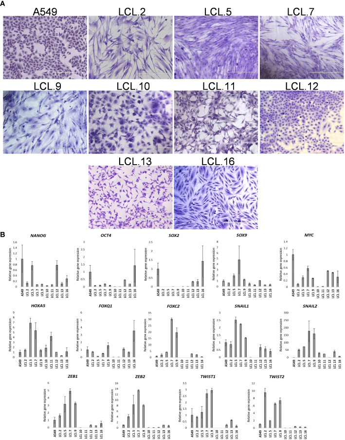
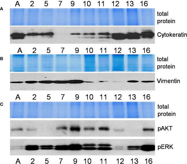
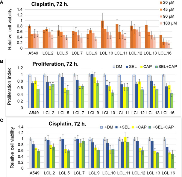
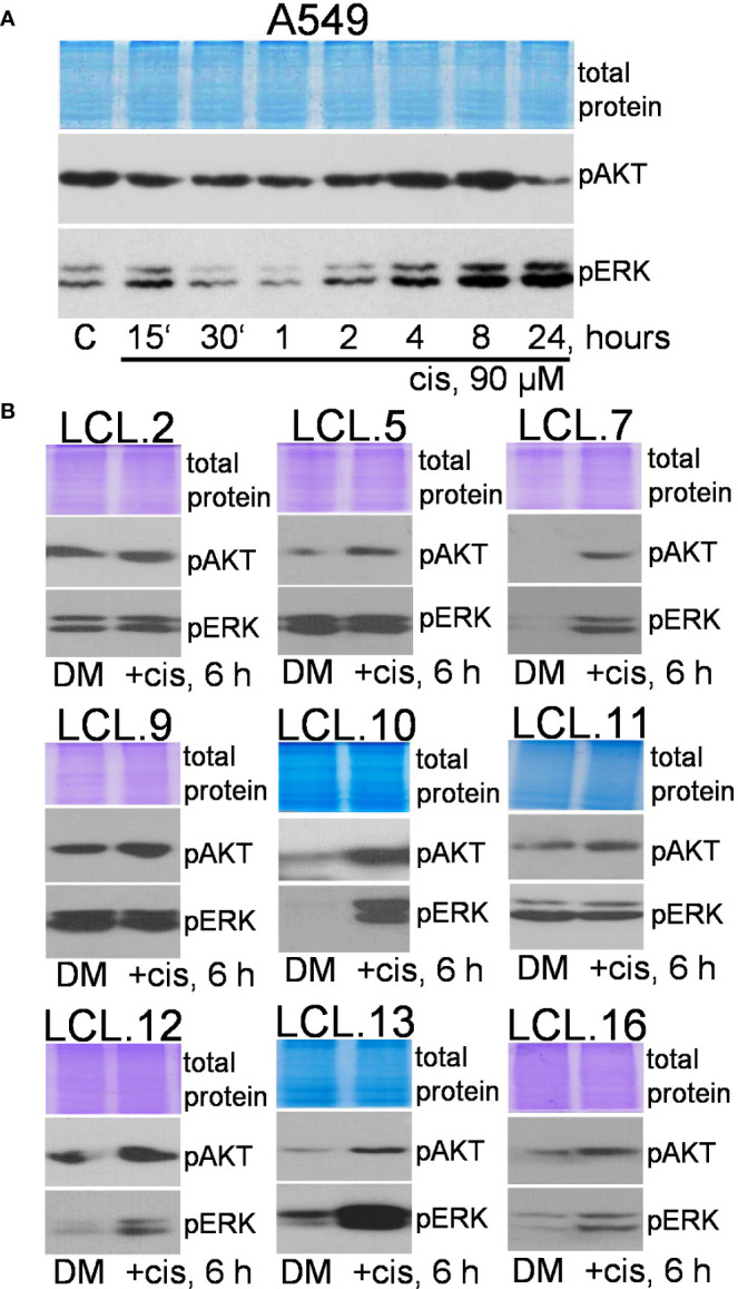
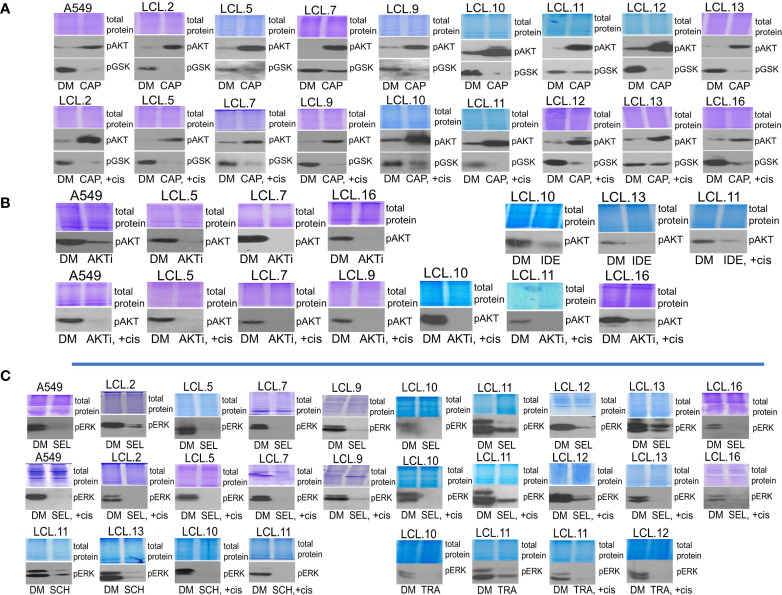
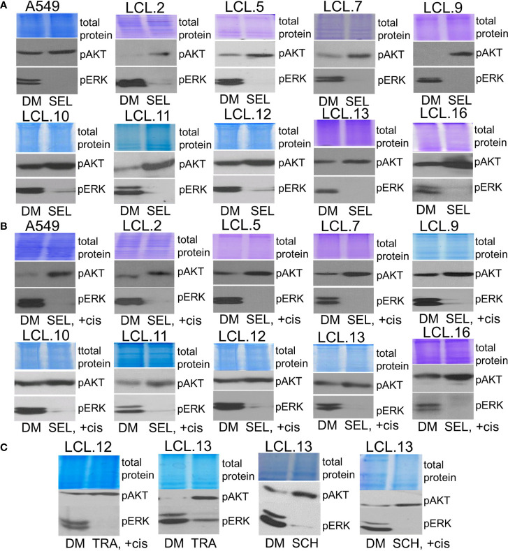
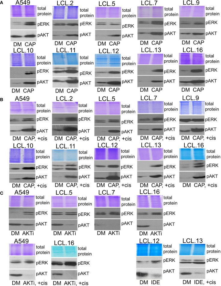
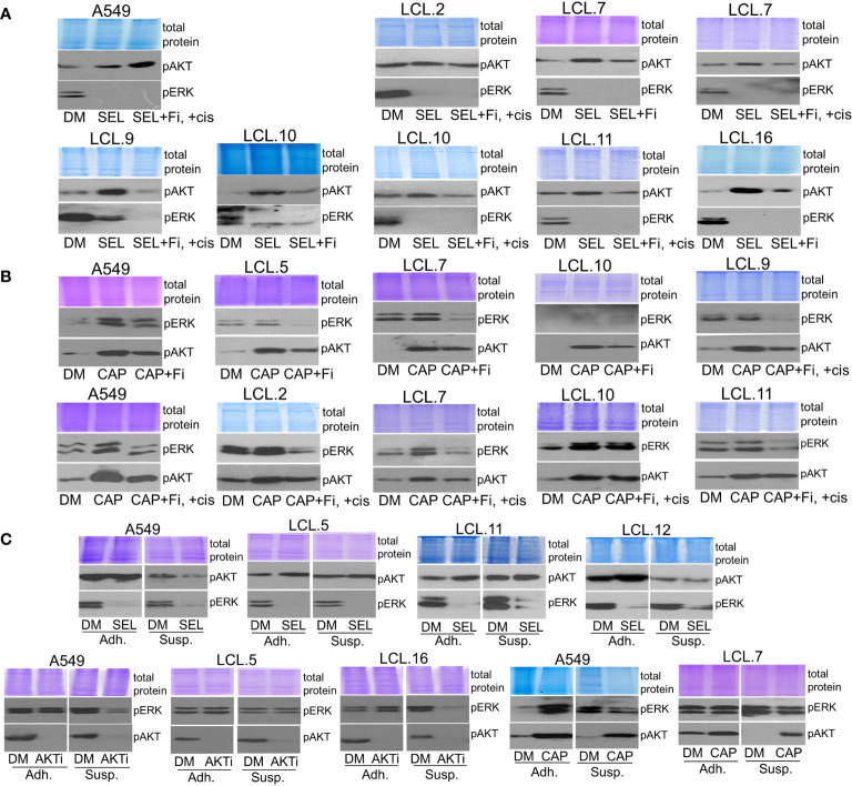
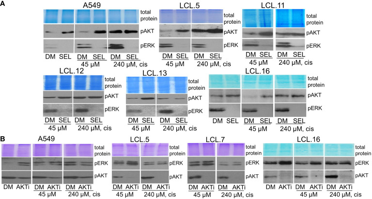
There is no doubt that cell signaling manipulation is a key strategy for anticancer therapy. Furthermore, cell state determines drug response. Thus, establishing the relationship between cell state and therapeutic sensitivity is essential for the development of cancer therapies. In the era of personalized medicine, the use of patient-derived ex vivo cell models is a promising approach in the translation of key research findings into clinics. Here, we were focused on the non-oncogene dependencies of cell resistance to anticancer treatments. Signaling-related mechanisms of response to inhibitors of MEK/ERK and PI3K/AKT pathways (regulators of key cellular functions) were investigated using a panel of patients' lung tumor-derived cell lines with various stemness- and EMT-related markers, varying degrees of ERK1/2 and AKT phosphorylation, and response to anticancer treatment. The study of interactions between kinases was the goal of our research. Although MEK/ERK and PI3K/AKT interactions are thought to be cell line-specific, where oncogenic mutations have a decisive role, we demonstrated negative feedback loops between MEK/ERK and PI3K/AKT signaling pathways in all cell lines studied, regardless of genotype and phenotype differences. Our work showed that various and distinct inhibitors of ERK signaling - selumetinib, trametinib, and SCH772984 - increased AKT phosphorylation, and conversely, inhibitors of AKT - capivasertib, idelalisib, and AKT inhibitor VIII - increased ERK phosphorylation in both control and cisplatin-treated cells. Interaction between kinases, however, was dependent on cellular state. The feedback between ERK and AKT was attenuated by the focal adhesion kinase inhibitor PF573228, and in cells grown in suspension, showing the possible role of extracellular contacts in the regulation of crosstalk between kinases. Moreover, studies have shown that the interplay between MEK/ERK and PI3K/AKT signaling pathways may be dependent on the strength of the chemotherapeutic stimulus. The study highlights the importance of spatial location of the cells and the strength of the treatment during anticancer therapy.

Keywords: AKT; ERK (extracellular signal-regulated kinase); cancer cell; cell signaling; cellular resistance; kinase inhibitors; lung cancer; targeted drugs.


Conflict of interest statement

The authors declare that the research was conducted in the absence of any commercial or financial relationships that could be construed as a potential conflict of interest.

Figures

Figure 1
Figure 1
Human NSCLC tumor-derived cell line characterization. (A) Morphology of cells from A549 and primary cell lines grown in vitro for more than 10 passages. Scale bars = 400 µm. Cells have been stained with crystal violet dye. (B) The relative expression of stem cell- and EMT-related transcription factors as determined by RT-qPCR (in respect to GAPDH). Data are presented as the mean ± SD (N = 2).
Figure 2
Figure 2
Characterization of cell lines (continued). Relative expression of mesenchymal and epithelial markers cytokeratin (A) and vimentin (B) (proteins of intermediate filaments family) and activated protein kinases phospho-ERK and phospho-AKT (C) as determined by the Western blot method. Representative pictures from more than 3 independent experiments are shown. Coomassie-stained polyacrylamide gels serve as loading controls. Numbers indicate cell lines as in Figure 1 ; the letter A stands for the cell line A549.
Figure 3
Figure 3
Impact of cisplatin and targeted drugs on cell viability of different lung cancer-derived cell lines. (A) Lung cancer-derived cells respond differently to cisplatin. Cell viability was determined by MTT assay and expressed in relation to the viability before the treatment (relative cell viability of initial control =1.0). DM – solvent DMSO. Data are expressed as means ± SD, N = 4. (B) Targeted drugs inhibit cell proliferation. Proliferation without inhibitors is normalized to 1.0. (C) Lung cancer cells respond differently to combinations of targeted drugs and conventional drug cisplatin. Cell survival without inhibitors is normalized to 1.0. Concentrations of cisplatin that induced 50% cell death after 72 h of treatment were used for each cell line. DM – vehicle control (DMSO), SEL – selumetinib (10 µM), CAP – capivasertib (10 µM).
Figure 4
Figure 4
Phosphorylation of ERK1/2 and AKT in lung cancer-derived cell lines after cisplatin treatment. (A) The dynamics of AKT and ERK phosphorylation in A549 cells. (B) Changes in AKT and ERK phosphorylation in lung tumor-derived cell lines after 6 hours of cisplatin treatment. Representative Western blots are shown. Coomassie-stained protein gels are presented as loading controls. The concentration of cisplatin was 90 µM.
Figure 5
Figure 5
Determining the efficacy of targeted drugs by phosphorylation of their targets. (A) Capivasertib, an AKT inhibitor, increases the phosphorylation of AKT itself. Capivasertib efficacy is demonstrated by inhibited phosphorylation of GSK3β, a downstream target of AKT. DM – vehicle control (DMSO), CAP – capivasertib, +cis – cisplatin. (B) AKT inhibitor VIII (AKTi) and PI3K inhibitor idelalisib (IDE) reduce AKT phosphorylation in control and cisplatin-treated cells. (C) MEK/ERK signaling inhibitors suppress ERK1/2 phosphorylation in control and cisplatin-treated cells. SEL – selumetinib, SCH – ERK inhibitor SCH772984, TRA – trametinib. Representative Western blots are shown. Coomassie-stained protein gels are presented as loading controls. 6-hour-long exposures to the drugs were used. The concentration of cisplatin was 90 µM, and that of inhibitors was 10 µM except for trametinib and SCH772984 (1 µM).
Figure 6
Figure 6
Crosstalk with negative feedback between ERK and AKT signaling pathways in human lung cancer-derived cell lines. (A, B) MEK/ERK pathway inhibitor selumetinib enhances AKT phosphorylation in the cell lines tested, control (A) and cisplatin-treated cells (B). (C) MEK inhibitor trametinib as well as ERK inhibitor SCH772984 increase AKT phosphorylation in control and in cisplatin-treated cells. Representative Western blots are shown. Coomassie-stained protein gels are presented as loading controls. 6-hour-long exposures to the drugs were used. DM – vehicle control (DMSO), SEL – selumetinib (10 µM), +cis – cisplatin (90 µM), TRA – trametinib (1 µM), SCH - SCH772984 (1 µM).
Figure 7
Figure 7
Crosstalk between ERK and AKT (continued). (A, B) AKT inhibitor capivasertib enhances ERK1/2 phosphorylation in control (A) and cisplatin-treated cells (B). (C) AKT inhibitor VIII enhances ERK1/2 phosphorylation in the cell lines tested, both control and cisplatin-treated cells. Similarly, PI3K inhibitor idelalisib increases ERK phosphorylation in human lung-derived cell lines. Representative Western blots are shown. Coomassie-stained protein gels are presented as loading controls. 6-hour-long exposures to the drugs were used. DM – vehicle control (DMSO), CAP – capivasertib (10 µM), +cis – cisplatin (90 µM), AKTi – AKT inhibitor VIII (10 µM), IDE – idelalisib (10 µM).
Figure 8
Figure 8
Dependence of the interplay between ERK and AKT signaling pathways on extracellular contacts. (A) Focal adhesion kinase inhibitor PF573228 (+Fi; 10 µM) attenuates the MEK kinase inhibitor selumetinib-induced increase of AKT phosphorylation in control and cisplatin-treated lung cancer-derived cells, except A549 cells. (B) PF573228 prevents the AKT inhibitor capivasertib-induced increase in ERK phosphorylation in control and cisplatin-treated lung cancer-derived cells. (C) Cells grown in suspension (Susp) under agitation, in contrast to adherent cells (Adh), do not show alternative kinase activation after the treatment with inhibitors. Representative Western blots are shown. Coomassie-stained protein gels are presented as loading controls. 6-hour-long exposures to the drugs were used. DM – vehicle control (DMSO), SEL – selumetinib (10 µM), +cis – cisplatin (90 µM), CAP – capivasertib (10 µM), AKTi – AKT inhibitor VIII (10 µM).
Figure 9
Figure 9
ERK and AKT crosstalk might be signal strength-dependent. Cells pretreated with a high concentration of cisplatin (240 µM) do not show alternative kinase AKT activation in response to ERK inhibition by selumetinib (A), nor ERK activation in response to AKT inhibitor VIII, contrary to untreated or low cisplatin concentration (45 µM)-treated cells (B). Representative Western blots are shown. Coomassie-stained protein gels are presented as loading controls. 6-hour-long exposures to the drugs were used. DM – vehicle control (DMSO), SEL – selumetinib (10 µM), cis - cisplatin, AKTi – AKT inhibitor VIII (10 µM).

Similar articles

Cited by

References

    1. Stulpinas A, Imbrasaitė A, Krestnikova N, Valerija Kalvelytė A. Recent approaches encompassing the phenotypic cell heterogeneity for anticancer drug efficacy evaluation. In: Tumor progression and metastasis. London, UK: IntechOpen; (2020). doi: 10.5772/intechopen.89395 - DOI
    1. Majeed U, Manochakian R, Zhao Y, Lou Y. Targeted therapy in advanced non-small cell lung cancer: current advances and future trends. J Hematol Oncol (2021) 14(1):108. doi: 10.1186/s13045-021-01121-2 - DOI - PMC - PubMed
    1. Mansoori B, Mohammadi A, Davudian S, Shirjang S, Baradaran B. The different mechanisms of cancer drug resistance: A brief review. Adv Pharm Bull (2017) 7(3):339–48. doi: 10.15171/apb.2017.041 - DOI - PMC - PubMed
    1. Panda M, Biswal BK. Cell signaling and cancer: A mechanistic insight into drug resistance. Mol Biol Rep (2019) 46(5):5645–59. doi: 10.1007/s11033-019-04958-6 - DOI - PubMed
    1. Lee S, Rauch J, Kolch W. Targeting MAPK signaling in cancer: Mechanisms of drug resistance and sensitivity. Int J Mol Sci (2020) 21(3):1102. doi: 10.3390/ijms21031102 - DOI - PMC - PubMed