

Review
doi: 10.1016/j.bjae.2022.11.008.
Epub 2022 Dec 28.
The cervical plexus
Affiliations
- PMID: 36686890
- PMCID: PMC9845551
- DOI: 10.1016/j.bjae.2022.11.008
Item in Clipboard
Review
The cervical plexus
BJA Educ.
2023 Feb.
No abstract available
Keywords: cervical plexus; cervical plexus block; nerve block.
Conflict of interest statement
The authors declare that they have no conflicts of interest.
Figures
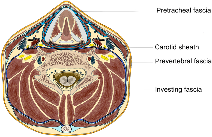
Axial cross section of the neck at C5 level demonstrating the layers of deep cervical fascia. The investing fascia is the most superficial, the pretracheal fascia is divided into muscular and visceral components and the prevertebral layer is the deepest.
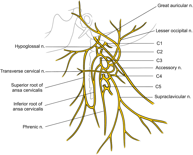
Branches of the cervical plexus. C5 does not contribute to the cervical plexus, but it is included to demonstrate the contribution to the phrenic nerve, and the accessory nerve is included to demonstrate its association to the plexus. n., nerve.
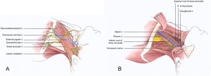
(A) The nerves of the superficial branches of the cervical plexus as they emerge from the posterior border of the sternocleidomastoid muscle. (B) The deep branches of the cervical plexus. The sternocleidomastoid is lifted in this illustration to allow visualisation of the deep branches of the plexus and demonstrate the close association to the hypoglossal, phrenic, vagus and spinal accessory nerves. First published in The Abbott Pocket Guide to Practical Peripheral Nerve Blockade. m., muscle; n., nerve; v., vein.
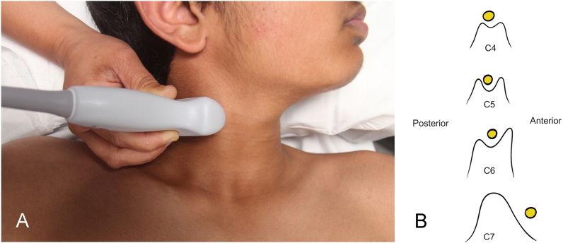
(A) Patient positioned for cervical plexus block. Note that the head is turned away and is resting on a surface so that the sternocleidomastoid muscle is relaxed. (B) Transverse processes of C4–7 with the position of the corresponding nerve root as visualised using ultrasound.
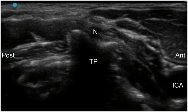
Ultrasound image of the C4 transverse process demonstrating C4 nerve root at the tip of the transverse process. ant, anterior; ICA, internal carotid artery; N, nerve; post, posterior; TP, transverse process.
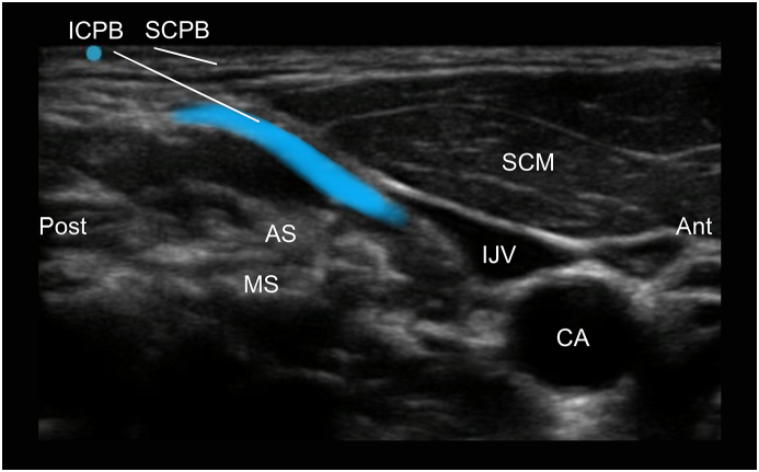
Ultrasound image of the anatomy relevant to the superficial (SCPB) and intermediate cervical plexus blocks (ICPB) at the C4 vertebral level. The intended spread of local anaesthetic within the ICPB plane is indicated by the blue area. Note that the external jugular vein is easily compressed by light pressure from the ultrasound probe as is not visible in this image. ant, anterior; AS, anterior scalene; CA, carotid artery; IJV, internal jugular vein; MS, middle scalene; post, posterior; SCM, sternocleidomastoid.
References
-
- Natale G., Condino S., Stecco A., et al. Is the cervical fascia an anatomical proteus? Surg Radiol Anat. 2015;37:1119–1127. - PubMed
-
- Kohan E.J., Wirth G.A. Anatomy of the neck. Clin Plast Surg. 2014;41:1–6. - PubMed
-
- Warshafsky D., Goldenberg D., Kanekar S.G. Imaging anatomy of deep neck spaces. Otolaryngol Clin North Am. 2012;45:1203–1221. - PubMed
-
- Guidera A.K., Dawes P.J., Fong A., Stringer M.D. Head and neck fascia and compartments: no space for spaces. Head Neck. 2014;36:1058–1068. - PubMed
-
- Caliot P., Dumont D., Bousquet V., Midy D. A note on the anastomoses between the hypoglossal nerve and the cervical plexus. Surg Radiol Anat. 1986;8:75–79. - PubMed
Publication types
LinkOut - more resources
Full Text Sources
