. 2023 Jan 13;12(1):2163781.
doi: 10.1080/2162402X.2022.2163781. eCollection 2023.

In vivo dynamics and anti-tumor effects of EpCAM-directed CAR T-cells against brain metastases from lung cancer

Affiliations

In vivo dynamics and anti-tumor effects of EpCAM-directed CAR T-cells against brain metastases from lung cancer

Tao Xu et al. Oncoimmunology. .

Abstract

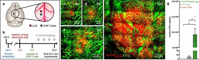
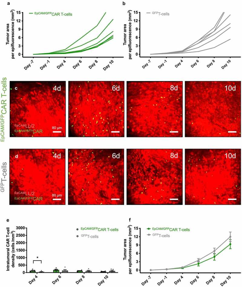
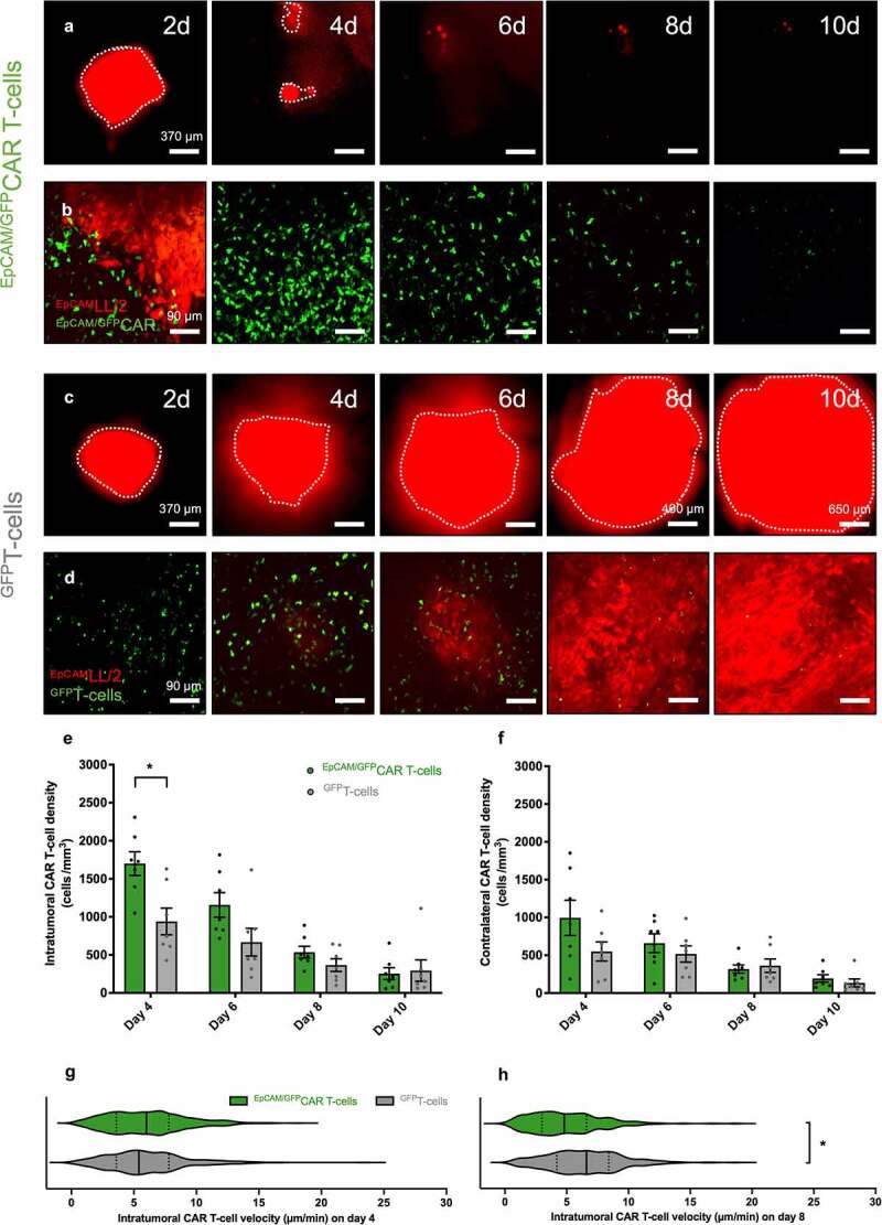
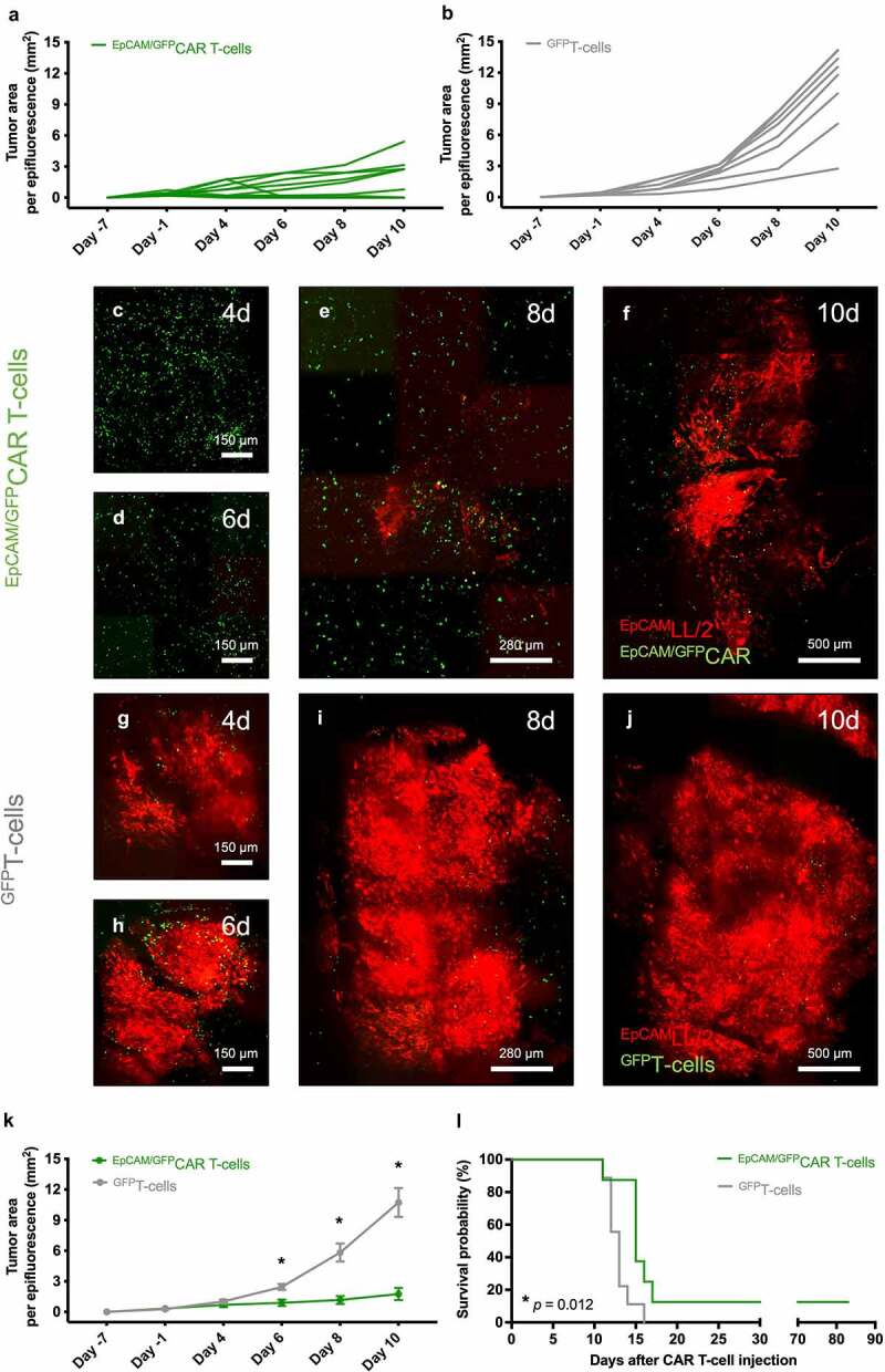
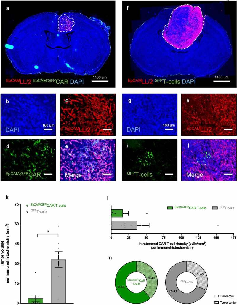
Lung cancer patients are at risk for brain metastases and often succumb to their intracranial disease. Chimeric Antigen Receptor (CAR) T-cells emerged as a powerful cell-based immunotherapy for hematological malignancies; however, it remains unclear whether CAR T-cells represent a viable therapy for brain metastases. Here, we established a syngeneic orthotopic cerebral metastasis model in mice by combining a chronic cranial window with repetitive intracerebral two-photon laser scanning-microscopy. This approach enabled in vivo-characterization of fluorescent CAR T-cells and tumor cells on a single-cell level over weeks. Intraparenchymal injection of Lewis lung carcinoma cells (expressing the tumor cell-antigen EpCAM) was performed, and EpCAM-directed CAR T-cells were injected either intravenously or into the adjacent brain parenchyma. In mice receiving EpCAM-directed CAR T-cells intravenously, we neither observed substantial CAR T-cell accumulation within the tumor nor relevant anti-tumor effects. Local CAR T-cell injection, however, resulted in intratumoral CAR T-cell accumulation compared to controls treated with T-cells lacking a CAR. This finding was accompanied by reduced tumorous growth as determined per in vivo-microscopy and immunofluorescence of excised brains and also translated into prolonged survival. However, the intratumoral number of EpCAM-directed CAR T-cells decreased during the observation period, pointing toward insufficient persistence. No CNS-specific or systemic toxicities of EpCAM-directed CAR T-cells were observed in our fully immunocompetent model. Collectively, our findings indicate that locally (but not intravenously) injected CAR T-cells may safely induce relevant anti-tumor effects in brain metastases from lung cancer. Strategies improving the intratumoral CAR T-cell persistence may further boost the therapeutic success.

Keywords: CAR T-cells; CNS tumor; adoptive immunotherapy; brain metastasis; histology; in vivo microscopy; lung cancer; survival.


Conflict of interest statement

Tao Xu, No disclosures; Philipp Karschnia, No disclosures; Bruno L. Cadilha, No disclosures; Sertac Dede, No disclosures; Michael Lorenz, No disclosures; Niklas Seewaldt, No disclosures; Elene Nikolaishvili, No disclosures; Katharina Müller, No disclosures; Jens Blobner, No disclosures; Nico Teske, No disclosures; Julika J. Herold, No disclosures; Kai Rejeski, Kite/Gilead: Research funding and travel support, Novartis: Honoraria, BMS/CELGENE: Consultancy, Honoraria; Sigrid Langer, No disclosures; Hannah Obeck, No disclosures; Theo Lorenzini, No disclosures; Matthias Mulazzani, No disclosures; Wenlong Zhang, No disclosures; Hellen Ishikawa-Ankerhold, No disclosures; Veit R. Buchholz, No disclosures; Marion Subklewe, No disclosures; Niklas Thon, No disclosures; Andreas Straube, No disclosures; Joerg-Christian Tonn, Research grants from Novocure and Munich Surgical Imaging, and Royalties from Springer Publisher Intl; Sebastian Kobold, S.K. has received honoraria from TCR2 Inc, Novartis, BMS and GSK, S.K. is an inventor of several patents in the field of immuno-oncology, S.K. received license fees from TCR2 Inc and Carina Biotech, S.K. received research support from TCR2 Inc. and Arcus Bioscience for work unrelated to the manuscript. Louisa von Baumgarten, No disclosures.

Figures

Figure 1.
Figure 1.
Experimental protocol and in vivo model of brain metastases from lung cancer. A: Position of the chronic cranial window (circle; diameter: 5.5 mm) and the injection sites of the tumor cells (red) as well as the locally injected (CAR) T-cells (green). For intravenous experiments, (CAR) T-cells were injected into the tail vein. The 3dBAR plugin of the Scalable Brain Atlas (Bakker et al. in Neuroinformatics, 2015) was used to create the panel, and the panel has been adapted from Zhang & Karschnia & von Mücke-Heim et al. (in Neoplasia, 2021). B: Schematic representation of the experimental design. C-F: Intracerebral growth of EpCAM/tdtLL/2 (red) following intraparenchymal tumor cell injection. Images represent mosaics of multiple maximum intensity projections with 400 µm depth from the brain surface. Blood vessels are highlighted via intravascular plasma staining using FITC-dextran (green). Note that the day count refers to the day after CAR T-cell injection (which is seven days after tumor injection). Scale bars: C, D: 200 µm; E: 275 µm; F: 333 µm. G: ELISA performed for murine IFN-γ on supernatants after co-culture of EpCAM/tdtLL/2 with naïve T-cells (orange), GFPT-cells (gray), or EpCAM/GFPCAR T-cells (green) for 24 hours (n = 4). Mean ± SEM. *p ≤ .05.
Figure 2.
Figure 2.
CAR T-cell density and anti-tumor effects after intravenous CAR T-cell injection. A, B: Individual tumor areas (mm2) measured by in vivo microscopy using epifluorescence on days −1, 4, 6, 8, and 10 after intravenous injection of EpCAM/GFPCAR T-cells (A; n = 6) or GFPT-cells (B; n = 6). C, D: Representative in vivo images of brain tumors (red) on days 4, 6, 8, and 10 after intravenous injection of EpCAM/GFPCAR T-cells (green; C) or GFPT-cells (green D) using two-photon laser scanning microscopy (maximum intensity projections with 400 µm depth from the brain surface). Note the slightly increased EpCAM/GFPCAR T-cell numbers on day 4 and the similar EpCAM/GFPCAR T-cell numbers during the following observation period. Scale bars: 80 µm. E: Density of intravenously injected (CAR) T-cells (cells/mm3) within the tumor on days 4, 6, 8, and 10 after receiving EpCAM/GFPCAR T-cells (green; n = 5) or GFPT-cells (gray; n = 5), as assessed by two-photon laser scanning microscopy. Mean ± SEM. *p ≤ 0.05. F: Pooled tumor areas (mm2) on days −1, 4, 6, 8, and 10 following intravenous injection of EpCAM/GFPCAR T-cells (green; n = 6) or GFPT-cells (gray; n = 6) determined by in vivo microscopy using epifluorescence. Mean ± SEM.
Figure 3.
Figure 3.
In vivo CAR T-cell dynamics after intraparenchymal injection. A-D: Representative in vivo images of brain tumors (red) on days 2, 4, 6, 8, and 10 after intraparenchymal injection of EpCAM/GFPCAR T-cells (green; A, B) or GFPT-cells (green; C, D) using epifluorescence (A, C; brain tumors delineated by dotted lines) and two-photon laser scanning microscopy (B, D; maximum intensity projections with 400 µm depth from the brain surface). Note the tumor regression and intratumoral CAR T-cell accumulation after local administration of EpCAM/GFPCAR T-cells. Scale bars: A, C: 370 µm (except GFPT-cells, day 8: 460 µm and day 10: 650 µm); B, D: 90 µm. E, F: Density of (CAR) T-cells (cells/mm3) within the tumor (e) and the healthy contralateral brain hemisphere (f) on days 4, 6, 8, and 10 after locally receiving EpCAM/GFPCAR T-cells (green; n = 7) or GFPT-cells (gray; n = 7), as assessed by two-photon laser scanning microscopy. Mean ± SEM. G, H: Intratumoral (CAR) T-cell velocity (µm/min) determined by two-photon laser scanning microscopy on day 4 (g) and day 8 (h) after local injection of EpCAM/GFPCAR T-cells (green; n = 7) or GFPT-cells (gray; n = 7). Straight lines in the violin plot indicate the median, dotted lines indicate quartiles. *p ≤ 0.05.
Figure 4.
Figure 4.
Tumor growth and survival after intraparenchymal injection of CAR T-cells. A, B: Individual tumor areas (mm2) measured by in vivo microscopy using epifluorescence on days −1, 4, 6, 8, and 10 after local injection of EpCAM/GFPCAR T-cells (A; n = 10) or GFPT-cells (B; n = 8). C-J: Brain tumor growth on days 4, 6, 8, and 10 after intraparenchymal injection of EpCAM/GFPCAR T-cells (c-f) or GFPT-cells (g-j) as illustrated by representative mosaics of multiple maximum intensity projections (with 400 µm depth from the brain surface) from two-photon laser scanning microscopy. EpCAM/GFPCAR T-cells or GFPT-cells are detected by their green fluorescent signal, and tumor cells are visualized based on their red fluorescent signal. Early in the observation period, EpCAM/GFPCAR T-cells were more evenly distributed throughout the tumor compared to GFPT-cells resulting in reduced tumor growth. Scale bars: C-D, G-H: 150 µm; E, I: 280 µm; F, J: 500 µm. K: Pooled tumor areas (mm2) on days −1, 4, 6, 8, and 10 following local injection of EpCAM/GFPCAR T-cells (green; n = 10) or GFPT-cells (gray; n = 8) determined by in vivo microscopy using epifluorescence. Mean ± SEM. *p ≤ 0.05. L: Kaplan–Meier estimates of survival in mice with brain tumors (injected seven days prior to (CAR) T-cell injection) following treatment with EpCAM/GFPCAR T-cells (green; n = 8) or GFPT-cells (gray; n = 9).
Figure 5.
Figure 5.
Immunofluorescence characterization of tumor growth and intratumoral CAR T-cells following intraparenchymal CAR T-cell injection. A-J: Histological sections of brains from mice with brain tumors excised 10 days after intraparenchymal injection of EpCAM/GFPCAR T-cells (a-e) or GFPT-cells (f-j). Sections were stained with an antibody against TdTomato to identify tumor cells (red), against GFP to visualize the (CAR) T-cell signal, and DAPI to allow detection of cell nuclei (blue). Tumors (dotted lines in A, F) were substantially smaller in mice which have received EpCAM/GFPCAR T-cells, whereas (CAR) T-cells were scattered through the tumor in scant numbers in both groups. B-E and G-J represent a selected intratumoral area from A and B, respectively. Scale bars: A, F: 1400 µm; B-E, G-J: 180 µm. K-M: Tumor volume (K; mm3), intratumoral (CAR) T-cell density (L; cells/mm), and distribution of intratumoral (CAR) T-cells (M; percentage) on day 10 after injection of EpCAM/GFPCAR T-cells (green; n = 9) or GFPT-cells (gray; n = 8) determined by immunofluorescence. Mean ± SEM. *p ≤ 0.05.

Similar articles

Cited by

References

    1. Achrol AS, Rennert RC, Anders C, Soffietti R, Ahluwalia MS, Nayak L, Peters S, Arvold ND, Harsh GR, Steeg PS, et al. Brain metastases. Nat Rev Dis Primers. 2019;5(1):5. doi: 10.1038/s41572-018-0055-y. - DOI - PubMed
    1. Shaw AT, Bauer TM, de Marinis F, Felip E, Goto Y, Liu G, Mazieres J, Kim D-W, Mok T, Polli A, et al. First-Line Lorlatinib or Crizotinib in Advanced ALK -Positive Lung Cancer. N Engl J Med. 2020;383(21):2018–13. doi: 10.1056/NEJMoa2027187. - DOI - PubMed
    1. Karschnia P, Le Rhun E, Vogelbaum MA, et al. The evolving role of neurosurgery for central nervous system metastases in the era of personalized cancer therapy. Eur J Cancer. 2021;156:93–108. doi: 10.1016/j.ejca.2021.07.032. - DOI - PubMed
    1. Goldberg SB, Schalper KA, Gettinger SN, Mahajan A, Herbst RS, Chiang AC, Lilenbaum R, Wilson FH, Omay SB, Yu JB, et al. Pembrolizumab for management of patients with NSCLC and brain metastases: long-term results and biomarker analysis from a non-randomised, open-label, phase 2 trial. Lancet Oncol. 2020;21(5):655–663. doi: 10.1016/S1470-2045(20)30111-X. - DOI - PMC - PubMed
    1. Chong EA, Ruella M, Schuster SJ.. Five-Year Outcomes for Refractory B-Cell Lymphomas with CAR T-Cell Therapy. N Engl J Med. 2021;384(7):673–674. doi: 10.1056/NEJMc2030164. - DOI - PubMed

Substances