

Review
doi: 10.1007/s13193-022-01591-x.
Epub 2022 Jul 16.
Imaging of the Infratemporal Fossa: a Comprehensive Pictorial Essay
Affiliations
- PMID: 36687258
- PMCID: PMC9845485
- DOI: 10.1007/s13193-022-01591-x
Item in Clipboard
Review
Imaging of the Infratemporal Fossa: a Comprehensive Pictorial Essay
Indian J Surg Oncol.
2022 Dec.
Abstract
While radiologists are familiar with the masticator space, the surgeons are more familiar with the infratemporal fossa (ITF). Though often used interchangeably, there exists a subtle difference between them, which needs to be understood. The close anatomical relationship of the infratemporal fossa to critical structures makes timely diagnosis vital. In this pictorial review, we present a spectrum of various pathologies affecting ITF.
Keywords: Anatomy; Computed Tomography; Infratemporal Fossa; Pathologies.
© The Author(s), under exclusive licence to Indian Association of Surgical Oncology 2022.
Conflict of interest statement
Conflict of InterestThe authors declare no competing interests.
Figures

Anatomy of infratemporal fossa. Axial (A) and coronal (B) CT sections show outline of the infratemporal fossa (yellow) and masticator space (purple)
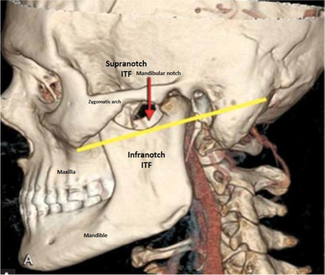
CT volume rendered technique reconstruction image showing mandibular notch as a demarcation line to classify as supranotch and infranotch compartment of ITF

CECT of paranasal sinuses of 36-year-old COVID patient with invasive fungal sinusitis. CECT axial (A) and coronal (B) reformatted images show soft tissue mucosal thickening in bilateral maxillary sinuses with extension into the infratemporal fossa via pterygopalatine fissure

CE-MRI of a 17-year-old male with juvenile nasal angiofibroma. Post-contrast T1 fat sat images reveal an avidly enhancing mass in the bilateral nasal cavity extending into the right infratemporal fossa with widened pterygomaxillary fissure

MRI of a 43-year- old male of trigeminal schwannoma. Axial T2-weighted images (A) show solid cystic lesion involving the right infratemporal fossa showing heterogenous enhancement on post-contrast images (B). It is involving posterolateral wall of maxillary wall

CT images of a 45-year-old male patient of Rosai Dorfman disease. CT images show soft tissue mass in the right infratemporal fossa (A) as well as intraorbital, extraconal mass in left orbit (B)

CECT of a 51-year-old female patient of right buccal carcinoma. CECT images show enhancing mass in the right buccal space extending into the infratemporal fossa

CECT of a 40-year-old female patient with lymphoma. CECT coronal reformatted images show a homogenous nodal mass on the right side of the neck, extending into the infratemporal fossa

CT of 4-year-old patient with rhabdomyosarcoma. CECT images reveals soft tissue mass epicentered in right infratemporal fossa. The medial and lateral pterygoid muscles are not seen separate from the mass

Metastasis/deposits involving ITF. A CT of 14-year-old child — known case of acute myeloid leukemia (AML) with granulocytic sarcoma. CECT image shows soft tissue deposit in the left infratemporal fossa extending into the inferior orbital fissure. B CECT images of a 7-year-old patient with adrenal neuroblastoma with a metastatic deposit in the infratemporal fossa. CECT images show enhancing soft tissue in the right infratemporal fossa extending into the extraconal compartment of right orbit

Primary jaw tumors involving ITF. A, B CECT images of 5-year-old male child with desmoplastic fibroma left hemimandible show lytic destruction of ramus and angle of mandible on the left side with associated soft tissue in the infratemporal fossa. C, D CECT images of a 10-year-old female patient of osteogenic sarcoma show lytic sclerotic destruction of the left roof of orbit with a large soft tissue component extending into the infratemporal fossa and left nasal cavity. E, F CT images of 14 year of a male patient with jaw PNET shows destruction of the right angle of the mandible with spiculated periosteal reaction and a large soft tissue component in the infratemporal fossa and right submandibular space
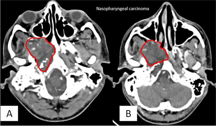
CT images of a 15-year-old male patient of nasopharyngeal carcinoma CT images show enhancing mass epicentered in nasopharynx on right side extending into ipsilateral infratemporal fossa via the pterygomaxillary fissure
References
-
- Jain S, Kumar A, Dhongade H, Varma R, Hathiram BT, Khattar VS. Imaging of parapharyngeal space and infratemporal fossa. Int J Otorhinolaryngol Clin. 2012;4(3):113–121. doi: 10.5005/jp-journals-10003-1096. - DOI
-
- Amit M, Bell D, Hunt PJ, Hanna E, Su SY, Kupferman M, Aashiq M, Takahashi H, Gidley PW, Nader ME, DeMonte F. Surgical management of carcinomas of the infratemporal fossa and skull base: patterns of failure and predictors of long-term outcomes. J Neurosurg. 2020;134(5):1392–1398. doi: 10.3171/2020.3.JNS192630. - DOI - PubMed
Publication types
LinkOut - more resources
Full Text Sources