

Monkeypox infection elicits strong antibody and B cell response against A35R and H3L antigens
- PMID: 36687315
- PMCID: PMC9838220
- DOI: 10.1016/j.isci.2023.105957
Monkeypox infection elicits strong antibody and B cell response against A35R and H3L antigens
Abstract
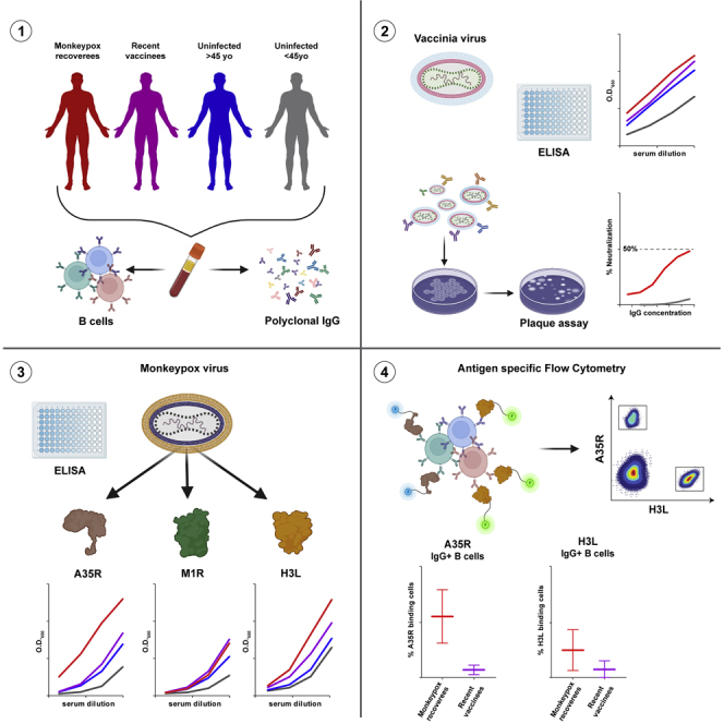
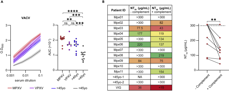
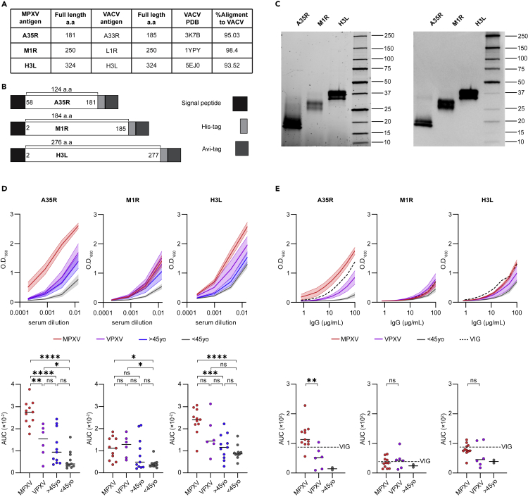
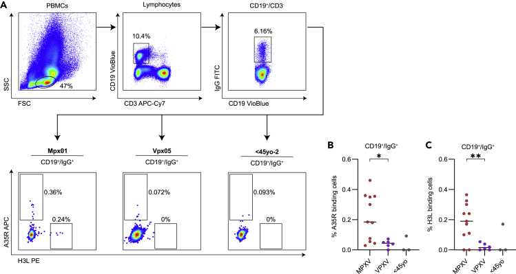
Monkeypox virus (MPXV) resides in two forms; mature and enveloped, and depending on it, distinct proteins are displayed on the viral surface. Here, we expressed two MPXV antigens from the mature, and one from the enveloped form, and tested their reactivity to sera of 11 MPXV recoverees while comparing to sera from recently and past vaccinated individuals. 8 out of 11 recoverees exhibited detectable neutralization levels against Vaccinia Lister. Sera from all recoverees bound strongly to A35R and H3L antigens. Moreover, the responses to A35R were significantly higher within the recoverees compared to both recently and past vaccinated donors. Lastly, A35R- and H3L-specific IgG+ B cells ranging from 0.03-0.46% and 0.11-0.36%, respectively, were detected in all recoverees (A35R), and in 9 out of 11 recoverees (H3L). Therefore, A35R and H3L represent MPXV immune targets and could be used in a heat-inactivated serological ELISA for the identification of recent MPXV infection.
Keywords: Immunological methods; Immunology; Virology.
© 2023 The Author(s).
Conflict of interest statement
The authors declare no conflict of interest.
Figures
Similar articles
-
Immunogenicity of monkeypox virus surface proteins and cross-reactive antibody responses in vaccinated and infected individuals: implications for vaccine and therapeutic development.Infect Dis Poverty. 2025 Feb 25;14(1):12. doi: 10.1186/s40249-025-01280-1. Infect Dis Poverty. 2025. PMID: 39994775 Free PMC article.
-
Development of multi-epitope vaccines against the monkeypox virus based on envelope proteins using immunoinformatics approaches.Front Immunol. 2023 Mar 13;14:1112816. doi: 10.3389/fimmu.2023.1112816. eCollection 2023. Front Immunol. 2023. PMID: 36993967 Free PMC article.
-
Multi-valent mRNA vaccines against monkeypox enveloped or mature viron surface antigens demonstrate robust immune response and neutralizing activity.Sci China Life Sci. 2023 Oct;66(10):2329-2341. doi: 10.1007/s11427-023-2378-x. Epub 2023 Jun 1. Sci China Life Sci. 2023. PMID: 37300753 Free PMC article.
-
Immunogenic proteins and potential delivery platforms for mpox virus vaccine development: A rapid review.Int J Biol Macromol. 2023 Aug 1;245:125515. doi: 10.1016/j.ijbiomac.2023.125515. Epub 2023 Jun 21. Int J Biol Macromol. 2023. PMID: 37353117 Free PMC article. Review.
-
Monkeypox virus: a re-emergent threat to humans.Virol Sin. 2022 Aug;37(4):477-482. doi: 10.1016/j.virs.2022.07.006. Epub 2022 Jul 9. Virol Sin. 2022. PMID: 35820590 Free PMC article. Review.
Cited by
-
Oral Infection, Oral Pathology and Salivary Diagnostics of Mpox Disease: Relevance in Dentistry and OMICs Perspectives.Int J Mol Sci. 2023 Sep 21;24(18):14362. doi: 10.3390/ijms241814362. Int J Mol Sci. 2023. PMID: 37762664 Free PMC article. Review.
-
Monkeypox virus H3L protein as the target antigen for developing neutralizing antibody and serological assay.Appl Microbiol Biotechnol. 2025 Apr 2;109(1):80. doi: 10.1007/s00253-025-13466-6. Appl Microbiol Biotechnol. 2025. PMID: 40172630 Free PMC article.
-
Identification of neutralizing nanobodies protecting against poxvirus infection.Cell Discov. 2025 Mar 25;11(1):31. doi: 10.1038/s41421-025-00771-7. Cell Discov. 2025. PMID: 40133273 Free PMC article.
-
Advances and challenges of mpox detection technology.Biosaf Health. 2024 Sep 11;6(5):260-269. doi: 10.1016/j.bsheal.2024.09.005. eCollection 2024 Oct. Biosaf Health. 2024. PMID: 40078738 Free PMC article. Review.
-
Monkeypox virus A29L protein as the target for specific diagnosis and serological analysis.Appl Microbiol Biotechnol. 2024 Nov 21;108(1):522. doi: 10.1007/s00253-024-13361-6. Appl Microbiol Biotechnol. 2024. PMID: 39570405 Free PMC article.
References
-
- OurWorldInData . 2022. Monkeypox Data Explorer.https://ourworldindata.org/explorers/monkeypox?tab=map&facet=none&Metric...
LinkOut - more resources
Full Text Sources
Other Literature Sources
Miscellaneous
