

Choroidal morphologic and vascular features in patients with unilateral idiopathic epiretinal membranes: An optical coherence tomography analysis integrated with assessment of retinal layers
- PMID: 36687460
- PMCID: PMC9853170
- DOI: 10.3389/fmed.2022.1083601
Choroidal morphologic and vascular features in patients with unilateral idiopathic epiretinal membranes: An optical coherence tomography analysis integrated with assessment of retinal layers
Abstract
Introduction: Integrated analysis of retinal and choroidal morphologic and vascular features is urgently needed to examine whether and how these two elements interact with each other, thus contributing to visual impairment in patients with idiopathic epiretinal membranes (iERMs).
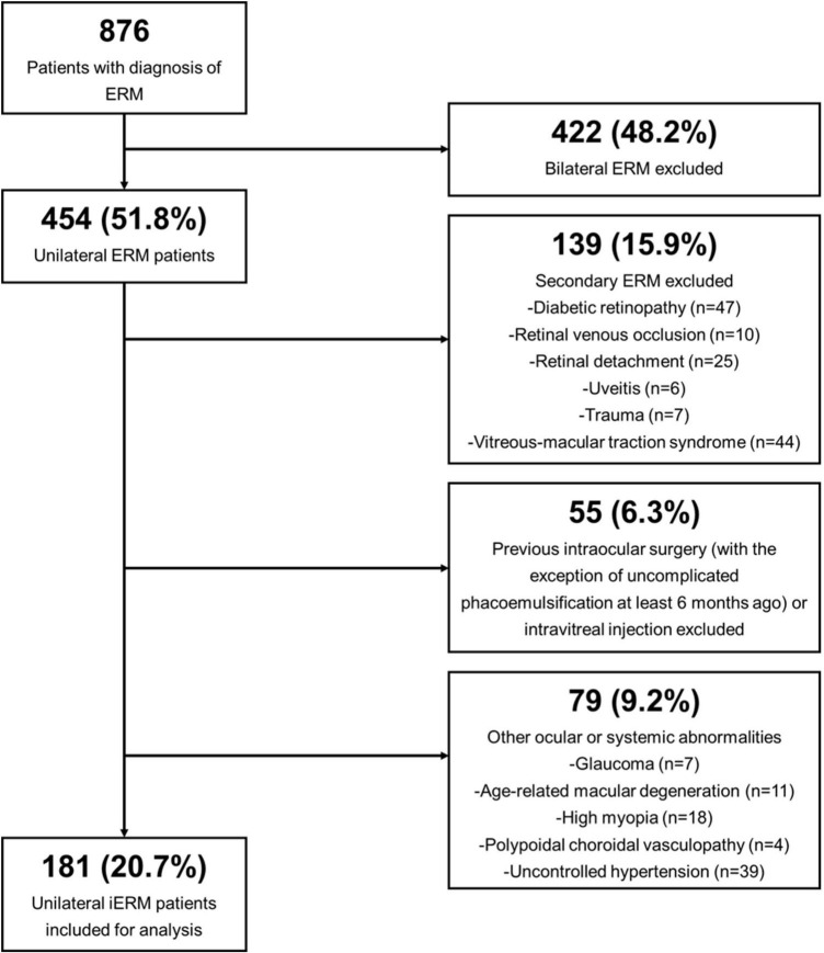
Methods: An observational retrospective study consisting of 181 patients diagnosed with unilateral iERM between August 2019 and July 2022 was carried out at Peking University Third Hospital. All patients underwent a standardized set of ophthalmologic examinations, including EDI-OCT and OCTA scanning, and were subsequently categorized into four stages according to current classification schemes based on their OCT findings. Altogether, 15 qualitative and quantitative parameters of both the retina (full-layer, inner and outer layers) and choroid were identified.
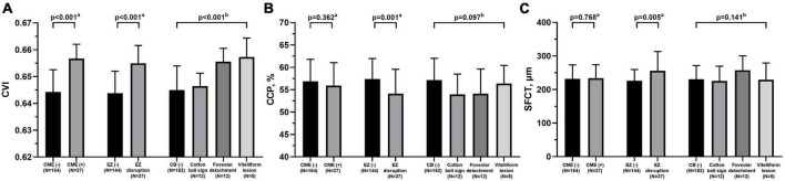
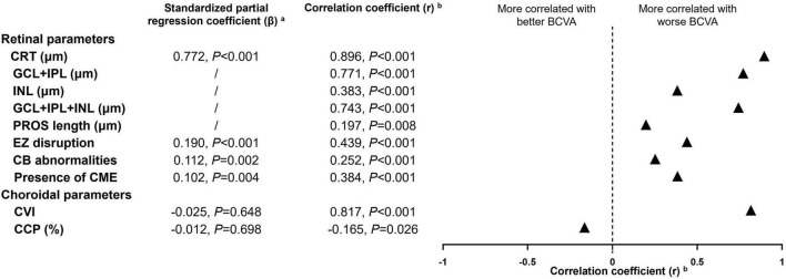
Results: The results revealed variations in the choroidal vascularity index (CVI) among different stages of iERMs (p < 0.001) for the first time. Distributions of retinal parameters across four stages of iERMs were validated. Correlation analysis between choroidal and retinal parameters showed that the CVI was associated with both inner and outer retinal morphologic biomarkers. Functional damage to retinal integrity was determined to be a strong contributor to visual acuity reduction in iERMs.
Discussion: This study complemented our present understanding of posterior segment structural and vascular alterations in iERMs.
Keywords: choroidal capillary perfusion; choroidal vascularity index; choroidal vasculature; enhanced depth imaging optical coherence tomography (EDI-OCT); idiopathic epiretinal membrane.
Copyright © 2023 Wang, Yang, Wang and Li.
Conflict of interest statement
The authors declare that the research was conducted in the absence of any commercial or financial relationships that could be construed as a potential conflict of interest.
Figures
Similar articles
-
Insights into the underlying choroid in different stages of idiopathic epiretinal membranes after Viteromacular surgery.Acta Ophthalmol. 2023 Jun;101(4):403-412. doi: 10.1111/aos.15295. Epub 2022 Nov 21. Acta Ophthalmol. 2023. PMID: 36408816
-
Gene expression analysis identifies two distinct molecular clusters of idiopatic epiretinal membranes.Biochim Biophys Acta Mol Basis Dis. 2020 Dec 1;1866(12):165938. doi: 10.1016/j.bbadis.2020.165938. Epub 2020 Aug 20. Biochim Biophys Acta Mol Basis Dis. 2020. PMID: 32827649
-
Is choroidal vascularity index a useful marker in different stages of idiopathic epiretinal membranes?Photodiagnosis Photodyn Ther. 2021 Mar;33:102110. doi: 10.1016/j.pdpdt.2020.102110. Epub 2020 Nov 23. Photodiagnosis Photodyn Ther. 2021. PMID: 33242656
-
[Pathophysiology of macular diseases--morphology and function].Nippon Ganka Gakkai Zasshi. 2011 Mar;115(3):238-74; discussion 275. Nippon Ganka Gakkai Zasshi. 2011. PMID: 21476310 Review. Japanese.
-
Choroidal Vascularity Index: An In-Depth Analysis of This Novel Optical Coherence Tomography Parameter.J Clin Med. 2020 Feb 21;9(2):595. doi: 10.3390/jcm9020595. J Clin Med. 2020. PMID: 32098215 Free PMC article. Review.
Cited by
-
Comparison of Functional, Structural, and Microvascular Features in Different Stages of Idiopathic Epiretinal Membrane.J Clin Med. 2024 May 29;13(11):3188. doi: 10.3390/jcm13113188. J Clin Med. 2024. PMID: 38892898 Free PMC article.
References
LinkOut - more resources
Full Text Sources