

Burning the candle at both ends: Intraretinal signaling of intrinsically photosensitive retinal ganglion cells
- PMID: 36687522
- PMCID: PMC9853061
- DOI: 10.3389/fncel.2022.1095787
Burning the candle at both ends: Intraretinal signaling of intrinsically photosensitive retinal ganglion cells
Abstract
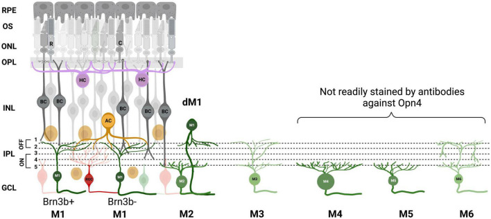
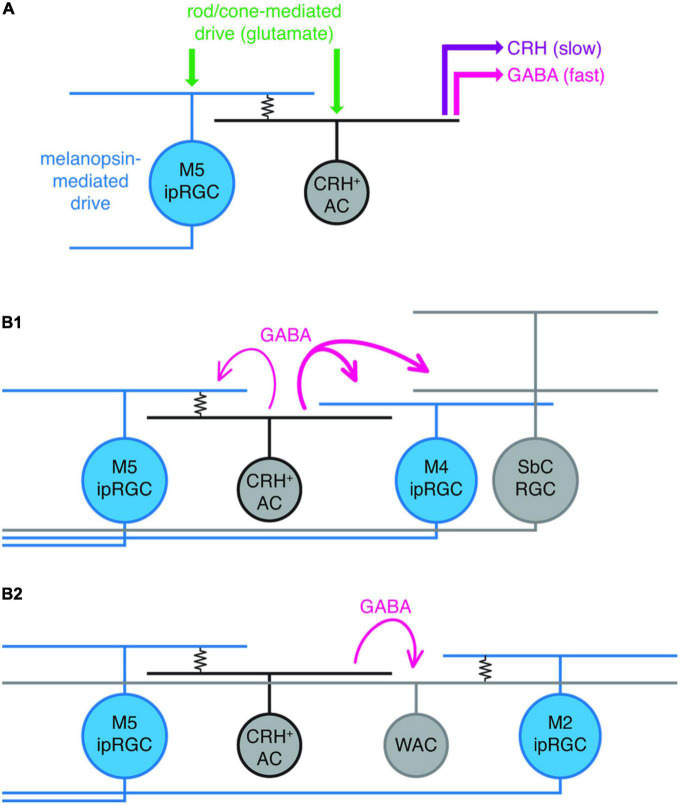
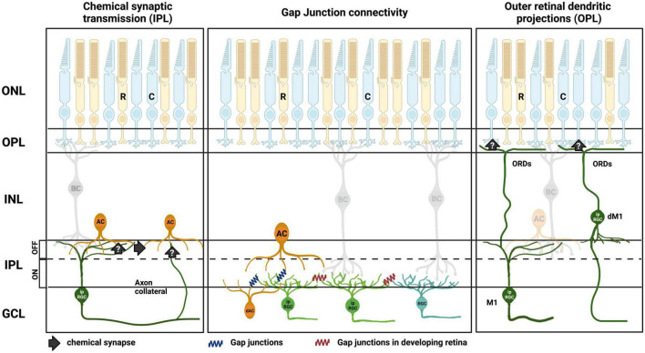
Intrinsically photosensitive retinal ganglion cells (ipRGCs) are photoreceptors located in the ganglion cell layer. They project to brain regions involved in predominately non-image-forming functions including entrainment of circadian rhythms, control of the pupil light reflex, and modulation of mood and behavior. In addition to possessing intrinsic photosensitivity via the photopigment melanopsin, these cells receive inputs originating in rods and cones. While most research in the last two decades has focused on the downstream influence of ipRGC signaling, recent studies have shown that ipRGCs also act retrogradely within the retina itself as intraretinal signaling neurons. In this article, we review studies examining intraretinal and, in addition, intraocular signaling pathways of ipRGCs. Through these pathways, ipRGCs regulate inner and outer retinal circuitry through both chemical and electrical synapses, modulate the outputs of ganglion cells (both ipRGCs and non-ipRGCs), and influence arrangement of the correct retinal circuitry and vasculature during development. These data suggest that ipRGC function plays a significant role in the processing of image-forming vision at its earliest stage, positioning these photoreceptors to exert a vital role in perceptual vision. This research will have important implications for lighting design to optimize the best chromatic lighting environments for humans, both in adults and potentially even during fetal and postnatal development. Further studies into these unique ipRGC signaling pathways could also lead to a better understanding of the development of ocular dysfunctions such as myopia.
Keywords: intraretinal; intrinsically photosensitive retinal ganglion cells (ipRGCs); melanopsin; retina; retinal processing.
Copyright © 2023 Raja, Milosavljevic, Allen and Cameron.
Conflict of interest statement
The authors declare that the research was conducted in the absence of any commercial or financial relationships that could be construed as a potential conflict of interest.
Figures
Similar articles
-
M1 ipRGCs Influence Visual Function through Retrograde Signaling in the Retina.J Neurosci. 2016 Jul 6;36(27):7184-97. doi: 10.1523/JNEUROSCI.3500-15.2016. J Neurosci. 2016. PMID: 27383593 Free PMC article.
-
Photoreceptive Ganglion Cells Drive Circuits for Local Inhibition in the Mouse Retina.J Neurosci. 2021 Feb 17;41(7):1489-1504. doi: 10.1523/JNEUROSCI.0674-20.2020. Epub 2021 Jan 4. J Neurosci. 2021. PMID: 33397711 Free PMC article.
-
A new method to quantify the human visual threshold from melanopsin sensitive ganglion cells.Front Cell Neurosci. 2023 Mar 23;17:1132230. doi: 10.3389/fncel.2023.1132230. eCollection 2023. Front Cell Neurosci. 2023. PMID: 37032840 Free PMC article.
-
[Intrinsically photosensitive retinal ganglion cells].Ophthalmologe. 2022 Apr;119(4):358-366. doi: 10.1007/s00347-021-01476-4. Epub 2021 Aug 4. Ophthalmologe. 2022. PMID: 34350494 Free PMC article. Review. German.
-
Melanopsin-expressing, Intrinsically Photosensitive Retinal Ganglion Cells (ipRGCs).2008 Aug 1 [updated 2016 Nov 2]. In: Kolb H, Fernandez E, Jones B, Nelson R, editors. Webvision: The Organization of the Retina and Visual System [Internet]. Salt Lake City (UT): University of Utah Health Sciences Center; 1995–. 2008 Aug 1 [updated 2016 Nov 2]. In: Kolb H, Fernandez E, Jones B, Nelson R, editors. Webvision: The Organization of the Retina and Visual System [Internet]. Salt Lake City (UT): University of Utah Health Sciences Center; 1995–. PMID: 21413413 Free Books & Documents. Review.
Cited by
-
Multimodality in the Collicular Pathway: Towards Compensatory Visual Processes.Cells. 2025 Apr 25;14(9):635. doi: 10.3390/cells14090635. Cells. 2025. PMID: 40358159 Free PMC article. Review.
-
Pupillary Light Reflex Reveals Melanopsin System Alteration in the Background of Myopia-26, the Female Limited Form of Early-Onset High Myopia.Invest Ophthalmol Vis Sci. 2024 Jul 1;65(8):6. doi: 10.1167/iovs.65.8.6. Invest Ophthalmol Vis Sci. 2024. PMID: 38958970 Free PMC article.
References
Publication types
LinkOut - more resources
Full Text Sources