Review
. 2023 Jan 17:11:tkac057.
doi: 10.1093/burnst/tkac057. eCollection 2023.

Epigenetic regulation of macrophage polarization in wound healing

Affiliations
Review

Epigenetic regulation of macrophage polarization in wound healing

Cheng Chen et al. Burns Trauma. .

Abstract

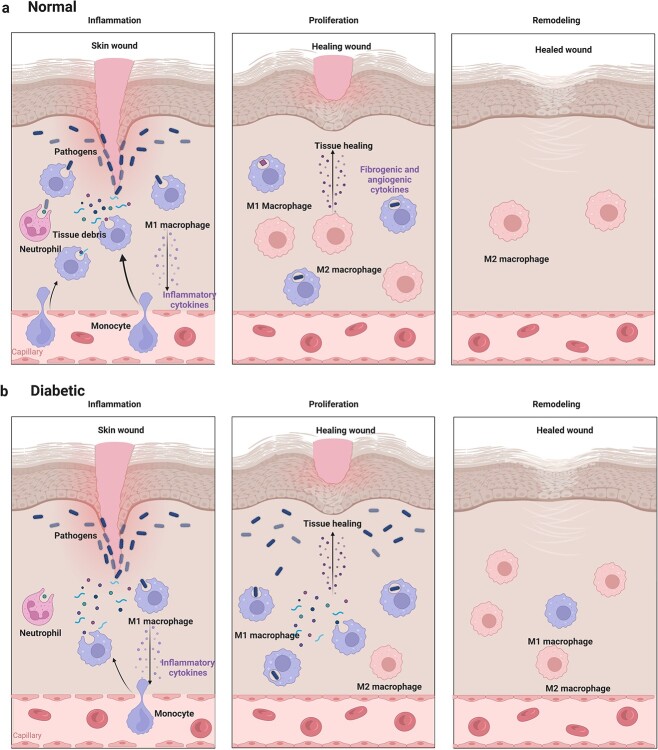
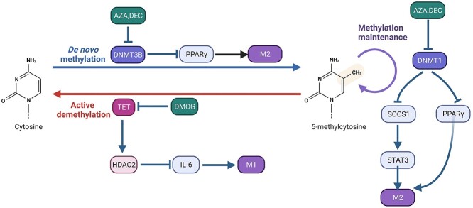
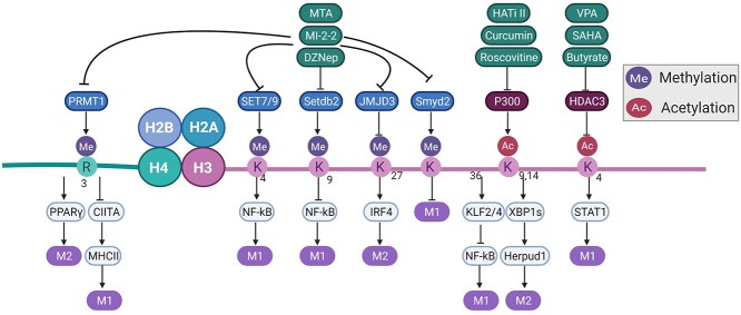
The immune microenvironment plays a critical role in regulating skin wound healing. Macrophages, the main component of infiltrating inflammatory cells, play a pivotal role in shaping the immune microenvironment in the process of skin wound healing. Macrophages comprise the classic proinflammatory M1 subtype and anti-inflammatory M2 population. In the early inflammatory phase of skin wound closure, M1-like macrophages initiate and amplify the local inflammatory response to disinfect the injured tissue. In the late tissue-repairing phase, M2 macrophages are predominant in wound tissue and limit local inflammation to promote tissue repair. The biological function of macrophages is tightly linked with epigenomic organization. Transcription factors are essential for macrophage polarization. Epigenetic modification of transcription factors determines the heterogeneity of macrophages. In contrast, transcription factors also regulate the expression of epigenetic enzymes. Both transcription factors and epigenetic enzymes form a complex network that regulates the plasticity of macrophages. Here, we describe the latest knowledge concerning the potential epigenetic mechanisms that precisely regulate the biological function of macrophages and their effects on skin wound healing.

Keywords: Epigenetics; Immune microenvironment; Macrophage polarization; Signaling pathways; Wound healing.


Figures

Figure 1
Figure 1
Macrophages in normal and diabetic wounds. (a) After injury, the recruitment of bone marrow-derived macrophages (BMDMs) plays an essential role in the inflammatory phase of wound healing. M1 macrophages clear pathogens and damaged tissues to avoid persistent stimulation by damage-associated molecular patterns and pathogen-associated molecular patterns and promote the transformation from inflammation to proliferation. At the remodeling stage, macrophages fade away [16]. (b) In diabetic wounds, delayed recruitment and short-lived BMDMs are caused by hyperglycemia, along with an attenuated ability to clear pathogens and damaged tissues [13]. The transformation from inflammation to proliferation and the disappearance of macrophages cannot occur in a timely manner
Figure 2
Figure 2
DNA methylation in regulating macrophage polarization. DNA methyltransferase is involved in controlling DNA methylation and downstream transcription factors. The inhibitors that regulate DNA methyltransferase are in green boxes. AZAazacytidine, DEC decitabine, DMOG dimethyloxallyl glycine, HDAC2 histone deacetylase2, TET ten-eleven translocation enzymes, DNMT DNA methyltransferases, PPAR γ peroxisome proliferator-activated receptors γ, SOCS1 socs suppressor of cytokine signaling 1, STAT3 signal transducers and activators of transcription 3
Figure 3
Figure 3
A portion of the histone modifications that participate in regulation of macrophage polarization. Histone modification enzymes involved in controlling macrophage polarization and downstream transcription factors. The modification site and modification type are shown on H3 and H4. The inhibitors that regulate histone modification are presented in green boxes. MTAmethylthioadenosine, MI-2-2 MLL–menin interaction inhibitor-2-2, DZNep 3-deazaneplanocin3, VPA valproic acid, SAHA suberoylanilide hydroxamic acid, HATi II histone acetyltransferase inhibitor II, SET7/9 Su(var)3–9, enhancer-of-zeste, trithorax7/9, SMYD SET and MYND domain containing, SETDB2 SET domain bifurcated histone lysine methyltransferase 2, JMJD3 Jumonji domain-containing 3, HDAC histone deacytelase, PRMT1 protein arginine methyltransferase 1, CIITAclass II transactivator, PPAR γ peroxisome proliferator-activated receptors γ, IRF4 IFN-regulatory factor 4, STATs signal transducers and activators of transcriptions, KLF2 Kruppel-like factors 2, XBP1s X-box protein 1, Herpud1 homocysteine inducible endoplasmic reticulum protein with ubiquitin-like domain 1

Similar articles

Cited by

References

    1. He L, Marneros AG. Macrophages are essential for the early wound healing response and the formation of a fibrovascular scar. Am J Pathol. 2013;182:2407–17. - PMC - PubMed
    1. Rodrigues M, Kosaric N, Bonham CA, Gurtner GC. Wound healing: a cellular perspective. Physiol Rev. 2019;99:665–706. - PMC - PubMed
    1. Kimball A, Schaller M, Joshi A, Davis FM, denDekker A, Boniakowski A, et al. . Ly6C(hi) blood monocyte/macrophage drive chronic inflammation and impair wound healing in diabetes mellitus. Arterioscler Thromb Vasc Biol. 2018;38:1102–14. - PMC - PubMed
    1. Cavalli G, Heard E. Advances in epigenetics link genetics to the environment and disease. Nature. 2019;571:489–99. - PubMed
    1. Álvarez-Errico D, Vento-Tormo R, Sieweke M, Ballestar E. Epigenetic control of myeloid cell differentiation, identity and function. Nat Rev Immunol. 2015;15:7–17. - PubMed