. 2022 Aug 9;9(12):nwac156.
doi: 10.1093/nsr/nwac156. eCollection 2022 Dec.

Development and transdifferentiation into inner hair cells require Tbx2

Affiliations

Development and transdifferentiation into inner hair cells require Tbx2

Zhenghong Bi et al. Natl Sci Rev. .

Abstract

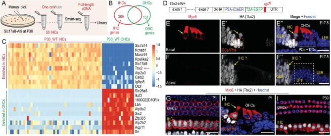
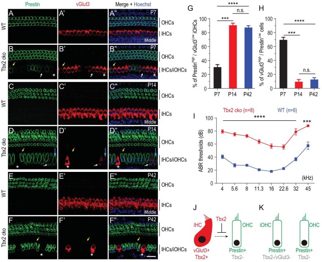
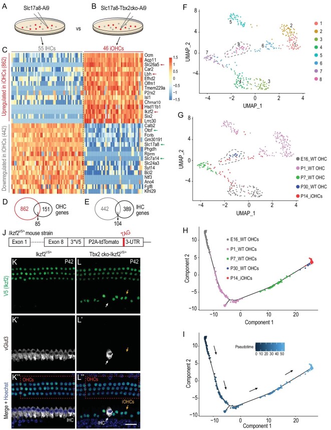
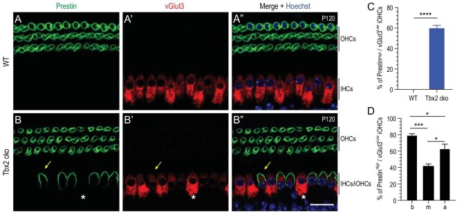
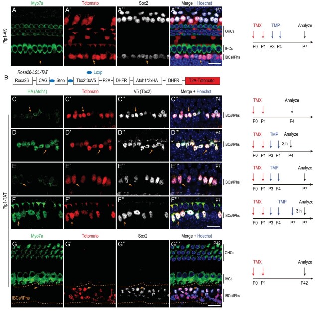
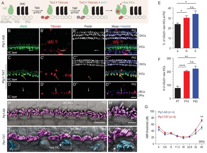
Atoh1 is essential for the development of both outer hair cells (OHCs) and inner hair cells (IHCs) in the mammalian cochlea. Whereas Ikzf2 is necessary for OHC development, the key gene required for IHC development remains unknown. We found that deletion of Tbx2 in neonatal IHCs led to their transdifferentiation into OHCs by repressing 26.7% of IHC genes and inducing 56.3% of OHC genes, including Ikzf2. More importantly, persistent expression of Tbx2 coupled with transient Atoh1 expression effectively reprogrammed non-sensory supporting cells into new IHCs expressing the functional IHC marker vGlut3. The differentiation status of these new IHCs was considerably more advanced than that previously reported. Thus, Tbx2 is essential for IHC development and co-upregulation of Tbx2 with Atoh1 in supporting cells represents a new approach for treating deafness related to IHC degeneration.

Keywords: Ikzf2; Tbx2; cochlea; inner hair cell; outer hair cell; supporting cell.


Figures

Figure 1.
Figure 1.
Tbx2 is highly expressed in IHCs but not OHCs. (A) and (B) Illustration of single-cell RNA-seq of adult IHCs at P30 by using smart-seq (A); 389 IHC genes and 151 OHC genes are identified (B). (C) Heat map showing top examples of differently expressed genes between IHCs and OHCs at P30. (D) Diagram of Tbx2*3 × HA-P2A-iCreER-T2A-EGFP/+ (Tbx2-HA/+); for details, please refer to Supplementary Fig. S1A–F. (E)–(I) Dual staining of HC marker Myo6 and HA (Tbx2) in cochlear samples from Tbx2-HA/+ mice at E17.5 (E)–(F’’), P1 (G) and (H) and P30 (I). Yellow arrows in (E)–(F’’): IHCs; dotted squares in (E’) and (H): IBCs/IPhs. IHCs, inner hair cells; OHCs, outer hair cells; GER, greater epithelial ridge cells; LER, lesser epithelial ridge cells; LPs, lateral progenitors; IBCs/IPhs, inner border cells/inner phalangeal cells; PCs, pillar cells; DCs, Deiters’ cells. Scale bar: 20 μm.
Figure 2.
Figure 2.
Loss of Tbx2 causes IHCs to gradually decrease vGlut3 but increase Prestin expression. (A)–(F’’) Dual whole-mount staining of vGlut3 and Prestin in cochlear samples from wild-type mice [WT; (A)–(A’’), (C)–(C’’) and (E)–(E’’)] and Tbx2 conditional knockout mice [Tbx2 cko; (B)–(B’’), (D)–(D’’) and (F)–(F’’)] at P7, P14 and P42, respectively. Yellow arrows: one PrestinHigh/vGlut3Low iOHC; white arrows: one vGlut3High/PrestinLow iOHC. Asterisks: IHCs that did not undergo cell-fate conversion; to simplify analysis, these cells were included in vGlut3High/PrestinLow population. (G) and (H) Quantification of PrestinHigh/vGlut3Low iOHCs (G) and vGlut3High/PrestinLow cells (H) in Tbx2 cko mice at P7, P14 and P42. Data are presented as means ± SEM. ***P < 0.001; ****P < 0.0001; n.s.: no significant difference. (I) ABR measurements between WT (blue line) and Tbx2 cko (red line) mice at P42; significantly different at all frequencies: ***P < 0.001; ****P < 0.0001. (J) and (K) Simplified model highlighting role of Tbx2 in IHC fate stabilization. After Tbx2 is deleted, IHCs become iOHCs (K). Scale bar: 20 μm.
Figure 3.
Figure 3.
Single-cell transcriptomic profiling of iOHCs. (A) and (B) Manual picking of P14_WT IHCs from Slc17a8-Ai9 mice (A) and P14_iOHCs from Slc17a8-Tbx2cko-Ai9 mice (B). (C) Transcriptomic comparison between P14_WT IHCs and P14_iOHCs. Relative to their expression in P14_WT IHCs, 862 and 442 genes are significantly upregulated and downregulated in P14_iOHCs, respectively. (D) and (E) Overlapping genes: 85 genes, including Slc26a5, Lbh and Ikzf2 (red arrows in C), overlap between the 862 upregulated and 151 defined OHC genes (D); and 104 genes, including Otof, Slc17a8 and Slc7a14 [green arrows in (C)], overlap between the 442 downregulated and 389 defined IHC genes. (F) and (G) UMAP analysis of cell mixtures covering E16_WT OHCs, P1_WT OHCs, P7_WT OHCs, P30_WT OHCs and P14_iOHCs; eight main cell clusters are revealed (F). Notably, 7/46 (15.2%) P14_iOHCs are assigned to Cluster 8 (gray dotted circle), which primarily comprises P30_WT OHCs. (H) and (I) Trajectory analysis of same cell mixtures as in (F) and (G). Arrows in (I): calculated developmental ages. (J) Simple illustration of how the Ikzf2V5/+ mouse strain is constructed. Briefly, three V5 tags are fused to Ikzf2 C-terminus; please also refer to Supplementary Fig. S5 for details. (K)–(L’’) Dual staining of V5 (Ikzf2) and vGlut3 in control Ikzf2V5/+(K)–(K’’) and Tbx2 cko -Ikzf2V5/+(L)–(L’’) cochleae at P42. Yellow arrows in (L)–(L’’): one iOHC expressing V5 (Ikzf2) but not vGlut3; white arrows in (L)–(L’’): one endogenous IHC not expressing V5 (Ikzf2) and maintaining vGlut3 expression. Scale bar: 20 μm.
Figure 4.
Figure 4.
Adult IHCs also transdifferentiate into OHCs when Tbx2 is conditionally deleted. (A)–(B’’) Double staining of Prestin and vGlut3 in WT (A)–(A’’) and Tbx2 cko (B)–(B’’) cochleae. Yellow arrows in (B)–(B’’): one iOHC with high Prestin expression but low vGlut3 expression; white asterisks: one suspected endogenous IHC maintaining high vGlut3 expression and not expressing Prestin. (C) Averaged percentage of PrestinHigh/vGlut3Low iOHCs in all turns of Tbx2 cko cochleae. Data are presented as means ± SEM. ****P < 0.0001. No iOHC is detected in WT cochleae. (D) Percentages of PrestinHigh/vGlut3Low iOHCs in basal (b), middle (m) and apical (a) turns of Tbx2 cko cochleae. Data are presented as means ± SEM. *P < 0.05; ***P < 0.001. Scale bar: 20 μm.
Figure 5.
Figure 5.
Successful transient Atoh1 and permanent Tbx2 ectopic expression in Plp1-TAT mice. (A)–(A’’’) Triple labeling for Myo7a, Tdtomato and Sox2 in control cochleae of Plp1-Ai9 mice first administered TMX (red arrows) and TMP (blue arrows) and then analysed at P7 (as depicted on the right). Orange arrows in (A)–(A’’’): one IBC/IPh that is Tdtomato+/Sox2+/Myo7a–. (B) Simple cartoon illustrating genetic model used to conditionally induce transient Atoh1 and permanent Tbx2 expression; please also refer to Supplementary Fig. S6 for details. (C)–(F’’’) Triple labeling for HA (Atoh1), Tdtomato and V5 (Tbx2) in Plp1-TAT mice subject to different treatments and analysed at distinct ages, as illustrated on the right. Orange arrows in (C)–(C’’’): one IBC/IPh with high Tdtomato and Tbx2 expression but weak Atoh1 expression. Orange arrows in (D)–(D’’’): one IBC/IPh with high Tdtomato and Tbx2 and also high Atoh1 expression, because Atoh1 is stabilized here by TMP treatment. Atoh1 expression is reversed to a weak level in Tdtomato+/Tbx2+ cells [orange arrows in (E)–(E’’’)] if mice are analysed at P7 (3 days after TMP treatment), but high expression can be restored in Tdtomato+/Tbx2+ cells [orange arrows in (F)–(F’’’)] if a third TMP treatment is administered 3 h before analysis at P7. (G)–(G’’’) Triple labeling for Myo7a, Tdtomato and Sox2 in cochleae of Plp1-TAT mice at P42. Arrows: one Tdtomato+/Sox2+ cell that does not express Myo7a and belongs to the IBCs/IPhs failing to become HCs. Scale bar: 20 μm (A’’’), (F’’’) and (G’’’).
Figure 6.
Figure 6.
Transient Atoh1 and permanent Tbx2 expression convert neonatal IBCs/IPhs into vGlut3+ new IHCs. (A) Simple cartoon illustrating key cellular events in reprogramming process. (B)–(D’’’) Triple labeling for IHC marker vGlut3, Tdtomato and OHC marker Prestin in control Plp1-Ai9 (B)–(B’’’) and experimental Plp1-TAT (C)–(D’’’) mice at P42. Images show visualization at HC layer (B)–(C’’’) or SC layer (D)–(D’’’). Arrows in (B)–(B’’’): one Tdtomato+ IBC/IPh that expresses neither vGlut3 nor Prestin. Arrows in (C)–(C’’’): one new IHC that is Tdtomato+/vGlut3+/Prestin–. Arrows in (D)–(D’’’): one Tdtomato+ IBC/IPh that fails to undergo cell-fate change and expresses neither vGlut3 nor Prestin. (E) Percentage of vGlut3+ new IHCs in three cochlear turns, basal (b), middle (m) and apical (a), of Plp1-TAT mice at P42. Data are presented as means ± SEM (n = 5). Percentage of new IHCs in apical turn is lower than in middle and basal turns (*P < 0.05). (F) Total numbers of vGlut3+ new IHCs in entire cochleae of Plp1-TAT mice at P7 (n = 3), P14 (n = 5) and P42 (n = 5). Data are presented as means ± SEM. **P < 0.01; ****P < 0.0001. (G) ABR measurements between Plp1-Ai9 (blue line) and Plp1-TAT (red line) mice at P42. No significant difference except at 45 kHz (**P < 0.01). (H)–(I’) SEM analysis of cochlear samples from control Plp1-Ai9 mice (H)–(H’) and experimental Plp1-TAT mice (I)–(I’). White arrows in (H)–(H’) and (I)–(I’): same endogenous IHCs; white arrowheads in (I)–(I’): same new IHCs. Scale bars: 2 μm (H’); 5 μm (H); 20 μm (D’’’).

Similar articles

Cited by

References

    1. Wu DK, Kelley MW. Molecular mechanisms of inner ear development. Cold Spring Harb Perspect Biol 2012; 4: a008409.10.1101/cshperspect.a008409 - DOI - PMC - PubMed
    1. Groves AK, Zhang KD, Fekete DM. The genetics of hair cell development and regeneration. Annu Rev Neurosci 2013; 36: 361–81.10.1146/annurev-neuro-062012-170309 - DOI - PMC - PubMed
    1. Montcouquiol M, Kelley MW. Development and patterning of the cochlea: from convergent extension to planar polarity. Cold Spring Harb Perspect Med 2020; 10: a033266.10.1101/cshperspect.a033266 - DOI - PMC - PubMed
    1. Bermingham NA, Hassan BA, Price SDet al. . Math1: an essential gene for the generation of inner ear hair cells. Science 1999; 284: 1837–41.10.1126/science.284.5421.1837 - DOI - PubMed
    1. Li S, Fan T, Li Cet al. . Fate-mapping analysis of cochlear cells expressing Atoh1 mRNA via a new Atoh1(3*HA-P2A-Cre) knockin mouse strain. Dev Dyn 2022; 251: 1156–7410.1002/dvdy.1453. - DOI - PubMed