

Enhanced phosphatidylserine exposure and erythropoiesis in Babesia microti-infected mice
- PMID: 36687590
- PMCID: PMC9846230
- DOI: 10.3389/fmicb.2023.1083467
Enhanced phosphatidylserine exposure and erythropoiesis in Babesia microti-infected mice
Erratum in
-
Erratum: Enhanced phosphatidylserine exposure and erythropoiesis in Babesia microti-infected mice.Front Microbiol. 2023 Feb 21;14:1157549. doi: 10.3389/fmicb.2023.1157549. eCollection 2023. Front Microbiol. 2023. PMID: 36896432 Free PMC article.
Abstract
Introduction: Babesia microti (B. microti) is the dominant species responsible for human babesiosis, which is associated with severe hemolytic anemia and splenomegaly because it infects mammalian erythrocytes. The actual prevalence of B. microti is thought to have been substantially underestimated.
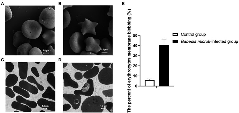
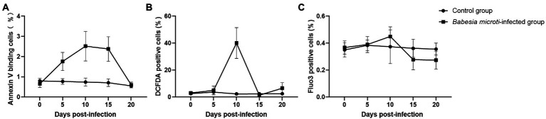
Methods: In this study, Bagg's albino/c (BALB/c) mice were intraperitoneally injected with B. microti-infected erythrocytes, and parasitemia was subsequently measured by calculating the proportion of infected erythrocytes. The ultrastructure of infected erythrocytes was observed using scanning and transmission electron microscopes. Quantifying phosphatidylserine (PS) exposure, oxidative stress, intracellular Ca2+, and erythropoiesis of erythrocytes were done using flow cytometry. The physiological indicators were analyzed using a Mindray BC-5000 Vet automatic hematology analyzer.
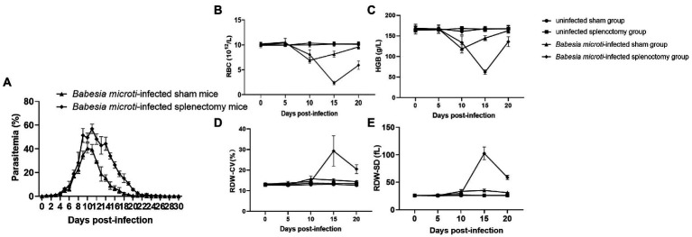
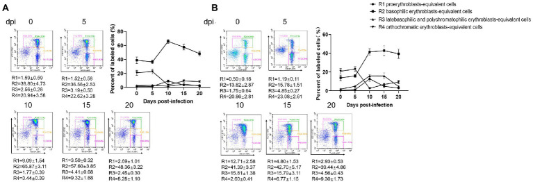
Results: Of note, 40.7 ± 5.9% of erythrocytes changed their structure and shrunk in the B. microti-infected group. The percentage of annexin V-positive erythrocytes and the levels of reactive oxygen species (ROS) in the erythrocytes were higher in the B. microti-infected group than in the control group at 10 dpi. Significant splenomegaly and severe anemia were also observed following B. microti infection. The parasitemia level in the B. microti-infected splenectomized group was higher than that of the B. microti-infected sham group. The population of early erythroblasts increased, and the late erythroblasts decreased in both the bone marrow and spleen tissues of the B. microti-infected group at 10 dpi.
Discussion: PS exposure and elevated ROS activities were hallmarks of eryptosis in the B. microti-infected group. This study revealed for the first time that B. microti could also induce eryptosis. At the higher parasitemia phase, the occurrence of severe anemia and significant changes in the abundance of erythroblasts in B. microti-infected mice group were established. The spleen plays a critical protective role in controlling B. microti infection and preventing anemia. B. microti infection could cause a massive loss of late erythroblasts and induce erythropoiesis.
Keywords: Babesia microti; babesiosis; eryptosis; erythrocyte; erythropoiesis.
Copyright © 2023 Song, Cai, Chen, Chen and Chen.
Conflict of interest statement
The authors declare that the research was conducted in the absence of any commercial or financial relationships that could be construed as a potential conflict of interest.
Figures
Similar articles
-
[Dynamics of routine blood tests in BALB/c mice with Babesia microti infection].Zhongguo Xue Xi Chong Bing Fang Zhi Za Zhi. 2018 Jun 20;30(3):300-306. doi: 10.16250/j.32.1374.2018119. Zhongguo Xue Xi Chong Bing Fang Zhi Za Zhi. 2018. PMID: 30019558 Chinese.
-
[Establishment of the experimental animal model of Babesia microti].Zhongguo Ji Sheng Chong Xue Yu Ji Sheng Chong Bing Za Zhi. 2012 Dec 30;30(6):423-7. Zhongguo Ji Sheng Chong Xue Yu Ji Sheng Chong Bing Za Zhi. 2012. PMID: 23484250 Chinese.
-
Temporal metabolic profiling of erythrocytes in mice infected with Babesia microti.Microb Pathog. 2023 Feb;175:105954. doi: 10.1016/j.micpath.2022.105954. Epub 2022 Dec 24. Microb Pathog. 2023. PMID: 36574865
-
Age-Related Differential Stimulation of Immune Response by Babesia microti and Borrelia burgdorferi During Acute Phase of Infection Affects Disease Severity.Front Immunol. 2018 Dec 7;9:2891. doi: 10.3389/fimmu.2018.02891. eCollection 2018. Front Immunol. 2018. PMID: 30619263 Free PMC article.
-
Investigating disease severity in an animal model of concurrent babesiosis and Lyme disease.Int J Parasitol. 2019 Feb;49(2):145-151. doi: 10.1016/j.ijpara.2018.06.006. Epub 2018 Oct 24. Int J Parasitol. 2019. PMID: 30367867 Free PMC article. Review.
Cited by
-
Apoptosis and eryptosis: similarities and differences.Apoptosis. 2024 Apr;29(3-4):482-502. doi: 10.1007/s10495-023-01915-4. Epub 2023 Nov 30. Apoptosis. 2024. PMID: 38036865 Review.
-
Temporal Dynamic Interplay of Mouse Proteome during Protozoan Babesia microti Infection Alone or with Borrelia burgdorferi Coinfection.J Proteome Res. 2025 Jul 4;24(7):3286-3299. doi: 10.1021/acs.jproteome.5c00015. Epub 2025 Jun 20. J Proteome Res. 2025. PMID: 40541258 Free PMC article.
-
Histopathological Aspects of the Influence of Babesia microti on the Placentas of Infected Female Rats.Vet Sci. 2024 Jan 3;11(1):18. doi: 10.3390/vetsci11010018. Vet Sci. 2024. PMID: 38250924 Free PMC article.
References
LinkOut - more resources
Full Text Sources
Miscellaneous