Review
. 2023 Jan 6:13:1083208.
doi: 10.3389/fmicb.2022.1083208. eCollection 2022.

Phage-encoded carbohydrate-interacting proteins in the human gut

Affiliations
Review

Phage-encoded carbohydrate-interacting proteins in the human gut

Daniela Rothschild-Rodriguez et al. Front Microbiol. .

Abstract

In the human gastrointestinal tract, the gut mucosa and the bacterial component of the microbiota interact and modulate each other to accomplish a variety of critical functions. These include digestion aid, maintenance of the mucosal barrier, immune regulation, and production of vitamins, hormones, and other metabolites that are important for our health. The mucus lining of the gut is primarily composed of mucins, large glycosylated proteins with glycosylation patterns that vary depending on factors including location in the digestive tract and the local microbial population. Many gut bacteria have evolved to reside within the mucus layer and thus encode mucus-adhering and -degrading proteins. By doing so, they can influence the integrity of the mucus barrier and therefore promote either health maintenance or the onset and progression of some diseases. The viral members of the gut - mostly composed of bacteriophages - have also been shown to have mucus-interacting capabilities, but their mechanisms and effects remain largely unexplored. In this review, we discuss the role of bacteriophages in influencing mucosal integrity, indirectly via interactions with other members of the gut microbiota, or directly with the gut mucus via phage-encoded carbohydrate-interacting proteins. We additionally discuss how these phage-mucus interactions may influence health and disease states.

Keywords: bacteriophage; glycans; glycosylation; gut; mucins; mucus; mucus-binding; mucus-degrading.


Conflict of interest statement

The authors declare that the research was conducted in the absence of any commercial or financial relationships that could be construed as a potential conflict of interest.

Figures
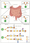
Figure 1
Figure 1
O-glycosylation of mucus mucins in the gut. (A) O-glycosylated mucins can contain 1-8 cores that stem off the protein backbone. Cores 1-4 make up the majority of O-glycosylated mucins in the gut. Cores 1 and 2 are found in the stomach and duodenum, core 3 is primarily found in the sigmoid colon and small intestine, and core 4 is localised to the colon. Some mucus-residing bacteria encode glycosyl hydrolases (GH) that cleave mucin glycans for bacteria to use as nutritional sources. This enzymatic breakdown of mucins is also part of the normal mucus turnover. The first N-acetylgalactosamine (GalNAc) of all cores is attached to the ser/thr residues via α-1 linkages which can be cleaved by the α-N-acetylgalactosaminidase, GH101. The cores then become differentiated by the addition of more groups to the initial GalNAc. Core 1 has galactose (Gal) attached to GalNAc via a β1-3 linkage, which can be cleaved by the β-galactosidases GH2, 20 and 42. Core 2 adds N-acetylglucosamine (GlcNAc) to the structure of core 1 via a β1-6 linkage to the GalNAc. The β1-6 and β1-3 bonds can be cleaved by GH2, 20 and 42. Core 3 includes a GlcNAc attached to GalNac via a β1-3 linkage, which is susceptible to cleavage by GH2, 20 and 42. Core 4 consists of two GlcNAc molecules bound to the GalNAc via a β1-6/β1-3 linkage, susceptible to cleavage by GH2, 20 and 42. Intestinal illustration taken from BioRender.com. FIGURE 1 (Continued)(B) Extension example of the glycan chain showing terminating glycans and additional enzymes involved in cleaving these. Gut mucin glycan chains are heavily sulfated (SO3), particularly at the distal ends of the intestinal tract, often terminating the chains to protect them from degradation. Sulfatases and sulfoglycosidases are non-GH-enzymes responsible for the desulfation of the chain to allow hydrolysis of the underlying glycans. Released sulfates can be reduced by sulfate-reducing bacteria and thus, their presence also promotes specific bacterial colonisation. Chains can also be terminated with fucose (Fuc) or sialic acid (Neu5Ac) residues that are thought to protect the underlying chain, while also serving as nutritional and energy sources for some bacteria. Neu5Ac is cleaved by sialidases (GH33) and Fuc by fucosidases (GH29 and 95).
Figure 2
Figure 2
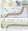
Phage-mucus interactions in the human gut. (A) In the small intestine, the microbiota can penetrate and reside in the single mucus layer, which is produced by goblet cells found within the epithelium layer. Phages can interact with and anchor to intestinal mucus in a suggested diffused gradient towards the lumen. This interaction allows the phage and host to be in close contact and co-exist. The mucus structure and function are defined by mucins, which are glycosylated with a high percentage of O-linked glycans. Some double-stranded DNA phages bind to mucus mucins via immunoglobulin-like (Ig-like) domains of the highly immunogenic outer capsid (Hoc) protein. Phage tail fibre proteins recognise glycans on the bacterial cell surface and thereby mediate phage adsorption to their host to initiate the infection process. Transmembrane mucins span the enterocyte lipid bilayer to form the glycocalyx. They consist of a cytoplasmic tail, protein backbone and mostly O-linked glycans. (B) The large intestine is comprised of an outer and inner mucus layer, but only the former is penetrable to the microbiota. Phages are suggested to co-exist with their hosts in this outer mucus layer in a similar diffused gradient to the small intestine. The dense, inner mucus layer of the large intestine consists of a tighter mesh of mucins with long branches of O-glycans that prevent bacteria-gut epithelium interactions in a healthy context.
Figure 3
Figure 3
Bacteriophage adherence to mucus (BAM) model expanded. (A) The mucus layer exists as a dynamic gradient with a higher density of glycoproteins closer to the epithelia, which is thought to cause a phage gradient within the mucus layer. The phage gradient consists of higher concentrations of phage closer to the epithelia, such that only continuous mucus secretion by goblet cells will push phages back out towards the lumen. Phage residence within the mucus layer allows them to be in close contact with their hosts as bacterial motility through the mucus increases their encounter rates; thus phage-bacterial interactions take place herein. (B) Potential phage-encoded carbohydrate active enzymes cleave mucin glycans allowing phages to diffuse down the mucus layer with more ease. (C–E) The phage gradient across the mucus layer is attributed to mucus density and phage diffusivity, with highest phage diffusivity occurring at lower mucin concentrations. It is also thought that different phages may have proteins with different binding and/or cleaving affinities to mucin glycans, affecting thus how deeply they can diffuse. (C) At <1% mucin concentration, Brownian motion is the dominating diffusion mode (high efficiency). (D) At ≥1% mucin concentration, the sub-diffusive continuous random time walk mode predominates (medium efficiency). (E) At 4% mucin concentration, the sub-diffusive fractional Brownian motion mode predominates (least efficient). Eventually phages will get stuck on the dense mucin mesh and only pushed out by continuous mucus secretion.
Figure 4
Figure 4
Disrupted mucus barrier and microbiome in Inflammatory Bowel Diseases. (A) At diagnosis, Crohn’s disease is most commonly located in the terminal ileum (45%), the colon (32%), ileocolonic (19%), and the upper gastrointestinal tract (4%; *). CD classically presents with discontinuous inflammation (skip lesions) of the gastrointestinal tract, where affected areas will see decreases in mucus production, loss of functional goblet cells, and increased inflammation whilst other sections appear healthy. Commensal bacterial richness and diversity are reduced while pathogenic bacteria and virulent dsDNA phage richness are increased. The mucosal barrier becomes weakened and ‘leaky’, consequently triggering inflammation, and can also lead to phage transcytosis into the lamina propria. (B) Inflammation in ulcerative colitis is localised to the colon, and it can continuously affect the large bowel at varying degrees. Virulent dsDNA phage richness, as well as sulfate-reducing, mucolytic and pro-inflammatory bacteria increase, while overall bacterial diversity and richness decrease. Mucus production, secretion and viscosity are substantially decreased, particularly in active disease, leading to the thinning of the mucus layer. This is attributed to the decreased presence of goblet cells in the epithelia, and the increased presence of immature goblet cells that secrete less and faulty mucus with decreased mucin glycosylation. It can also be partly attributed to the increased enzymatic activity of mucolytic bacteria. Eventually, the microbiota can penetrate the inner mucus layer, and worsen inflammatory responses.

Similar articles

Cited by

References

    1. Alemao C. A., Budden K. F., Gomez H. M., Rehman S. F., Marshall J. E., Shukla S. D., et al. (2021). Impact of diet and the bacterial microbiome on the mucous barrier and immune disorders. Allergy 76, 714–734. doi: 10.1111/ALL.14548, PMID: - DOI - PubMed
    1. Almeida G. M. F., Laanto E., Ashrafi R., Sundberg L.-R. (2019). Bacteriophage adherence to mucus mediates preventive protection against pathogenic bacteria. mBio 10:e01984-19. doi: 10.1128/MBIO.01984-19, PMID: - DOI - PMC - PubMed
    1. Bansil R., Turner B. S. (2018). The biology of mucus: composition, synthesis and organization. Adv Drug Del Rev 124, 3–15. doi: 10.1016/J.ADDR.2017.09.023, PMID: - DOI - PubMed
    1. Baquero F., Nombela C. (2012). The microbiome as a human organ. Clin. Microbiol. Infect. 18, 2–4. doi: 10.1111/J.1469-0691.2012.03916.X - DOI - PubMed
    1. Barr J. J., Auro R., Furlan M., Whiteson K. L., Erb M. L., Pogliano J., et al. (2013). Bacteriophage adhering to mucus provide a non–host-derived immunity. Proc. Natl. Acad. Sci. U. S. A. 110, 10771–10776. doi: 10.1073/PNAS.1305923110, PMID: - DOI - PMC - PubMed

LinkOut - more resources