

Role of advanced imaging techniques in the evaluation of oncological therapies in patients with colorectal liver metastases
- PMID: 36688023
- PMCID: PMC9850941
- DOI: 10.3748/wjg.v29.i3.521
Role of advanced imaging techniques in the evaluation of oncological therapies in patients with colorectal liver metastases
Abstract
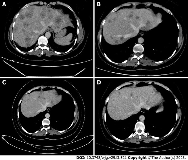
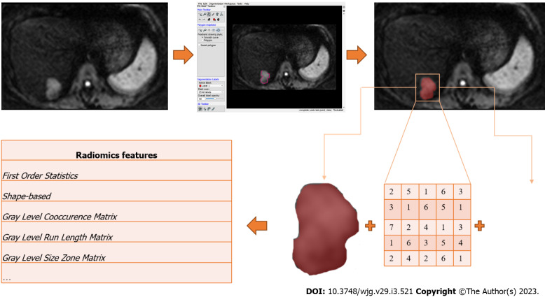
In patients with colorectal liver metastasis (CRLMs) unsuitable for surgery, oncological treatments, such as chemotherapy and targeted agents, can be performed. Cross-sectional imaging [computed tomography (CT), magnetic resonance imaging (MRI), 18-fluorodexoyglucose positron emission tomography with CT/MRI] evaluates the response of CRLMs to therapy, using post-treatment lesion shrinkage as a qualitative imaging parameter. This point is critical because the risk of toxicity induced by oncological treatments is not always balanced by an effective response to them. Consequently, there is a pressing need to define biomarkers that can predict treatment responses and estimate the likelihood of drug resistance in individual patients. Advanced quantitative imaging (diffusion-weighted imaging, perfusion imaging, molecular imaging) allows the in vivo evaluation of specific biological tissue features described as quantitative parameters. Furthermore, radiomics can represent large amounts of numerical and statistical information buried inside cross-sectional images as quantitative parameters. As a result, parametric analysis (PA) translates the numerical data contained in the voxels of each image into quantitative parameters representative of peculiar neoplastic features such as perfusion, structural heterogeneity, cellularity, oxygenation, and glucose consumption. PA could be a potentially useful imaging marker for predicting CRLMs treatment response. This review describes the role of PA applied to cross-sectional imaging in predicting the response to oncological therapies in patients with CRLMs.
Keywords: Colorectal cancer metastases; Computed tomography; Magnetic resonance imaging; Parametric imaging; Positron emission tomography; Prediction response.
©The Author(s) 2023. Published by Baishideng Publishing Group Inc. All rights reserved.
Conflict of interest statement
Conflict-of-interest statement: All the authors report no relevant conflicts of interest for this article.
Figures
Similar articles
-
Contrast-enhanced ultrasound can guide the therapeutic strategy by improving the detection of colorectal liver metastases.J Hepatol. 2021 Feb;74(2):419-427. doi: 10.1016/j.jhep.2020.09.036. Epub 2020 Oct 14. J Hepatol. 2021. PMID: 33065168
-
Colorectal cancer: Parametric evaluation of morphological, functional and molecular tomographic imaging.World J Gastroenterol. 2019 Sep 21;25(35):5233-5256. doi: 10.3748/wjg.v25.i35.5233. World J Gastroenterol. 2019. PMID: 31558870 Free PMC article. Review.
-
Liver metastases from colorectal cancer treated with conventional and antiangiogenetic chemotherapy: evaluation with liver computed tomography perfusion and magnetic resonance diffusion-weighted imaging.J Comput Assist Tomogr. 2011 Nov-Dec;35(6):690-6. doi: 10.1097/RCT.0b013e318230d905. J Comput Assist Tomogr. 2011. PMID: 22082538
-
Qualitative and quantitative parameters on hepatobiliary phase of gadoxetic acid-enhanced MR imaging for predicting pathological response to preoperative systemic therapy in colorectal liver metastases.Eur J Radiol. 2022 Dec;157:110572. doi: 10.1016/j.ejrad.2022.110572. Epub 2022 Oct 28. Eur J Radiol. 2022. PMID: 36327859
-
Non-invasive diagnostic imaging of colorectal liver metastases.World J Radiol. 2015 Jul 28;7(7):157-69. doi: 10.4329/wjr.v7.i7.157. World J Radiol. 2015. PMID: 26217455 Free PMC article. Review.
Cited by
-
Multiparametric MRI for characterization of the tumour microenvironment.Nat Rev Clin Oncol. 2024 Jun;21(6):428-448. doi: 10.1038/s41571-024-00891-1. Epub 2024 Apr 19. Nat Rev Clin Oncol. 2024. PMID: 38641651 Review.
-
Liver metastases: The role of magnetic resonance imaging.World J Gastroenterol. 2023 Sep 28;29(36):5180-5197. doi: 10.3748/wjg.v29.i36.5180. World J Gastroenterol. 2023. PMID: 37901445 Free PMC article. Review.
-
Immune checkpoint inhibitor-induced cholangitis-a three-case series.Explor Target Antitumor Ther. 2024;5(4):818-825. doi: 10.37349/etat.2024.00250. Epub 2024 Jul 19. Explor Target Antitumor Ther. 2024. PMID: 39280251 Free PMC article.
References
-
- Lam VW, Spiro C, Laurence JM, Johnston E, Hollands MJ, Pleass HC, Richardson AJ. A systematic review of clinical response and survival outcomes of downsizing systemic chemotherapy and rescue liver surgery in patients with initially unresectable colorectal liver metastases. Ann Surg Oncol. 2012;19:1292–1301. - PubMed
-
- Messersmith WA. NCCN Guidelines Updates: Management of Metastatic Colorectal Cancer. J Natl Compr Canc Netw. 2019;17:599–601. - PubMed
Publication types
MeSH terms
LinkOut - more resources
Full Text Sources
Medical
Research Materials
