. 2023 Jul;43(7):1142-1152.
doi: 10.1177/0271678X231152549. Epub 2023 Jan 23.

Noradrenaline released from locus coeruleus axons contracts cerebral capillary pericytes via α2 adrenergic receptors

Affiliations

Noradrenaline released from locus coeruleus axons contracts cerebral capillary pericytes via α2 adrenergic receptors

Nils Korte et al. J Cereb Blood Flow Metab. 2023 Jul.

Abstract

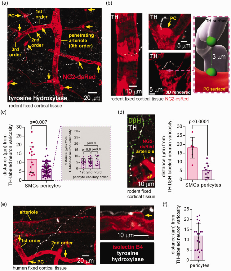
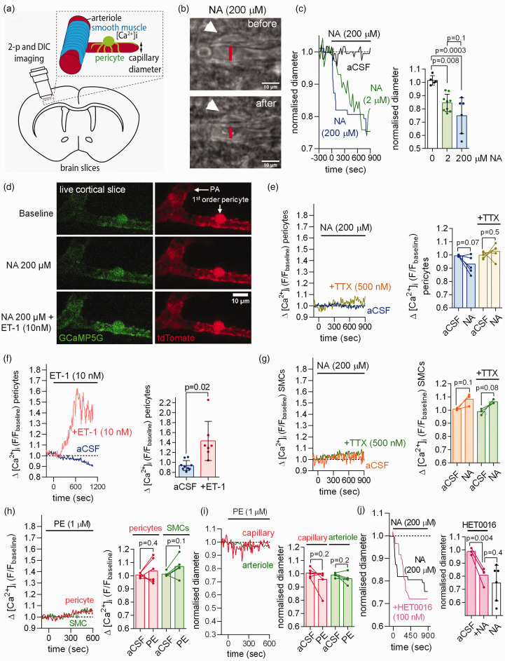
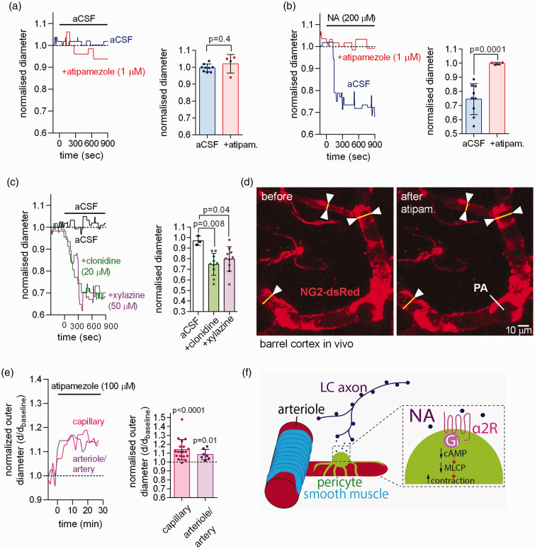
Noradrenaline (NA) release from locus coeruleus axons generates vascular contractile tone in arteriolar smooth muscle and contractile capillary pericytes. This tone allows neuronal activity to evoke vasodilation that increases local cerebral blood flow (CBF). Much of the vascular resistance within the brain is located in capillaries and locus coeruleus axons have NA release sites closer to pericytes than to arterioles. In acute brain slices, NA contracted pericytes but did not raise the pericyte cytoplasmic Ca2+ concentration, while the α1 agonist phenylephrine did not evoke contraction. Blocking α2 adrenergic receptors (α2Rs, which induce contraction by inhibiting cAMP production), greatly reduced the NA-evoked pericyte contraction, whereas stimulating α2Rs using xylazine (a sedative) or clonidine (an anti-hypertensive drug) evoked pericyte contraction. Noradrenaline-evoked pericyte contraction and capillary constriction are thus mediated via α2Rs. Consequently, α2Rs may not only modulate CBF in health and pathological conditions, but also contribute to CBF changes evoked by α2R ligands administered in research, veterinary and clinical settings.

Keywords: Noradrenaline; cAMP; calcium; locus coeruleus; pericyte.


Conflict of interest statement

The author(s) declared no potential conflicts of interest with respect to the research, authorship, and/or publication of this article.

Figures

Figure 1.
Figure 1.
Tyrosine hydroxylase- and DβH-labelled axons are in close proximity to rodent and human pericytes in the cerebral cortex. (a) Maximum intensity projection confocal image of tyrosine hydroxylase (TH) labelled axon varicosities (white, potential LC axon transmitter release sites) and pericytes (PC, yellow arrows) and arteriole smooth muscle cells (SMCs) labelled using dsRed under the NG2 promoter in the cortex of an adult NG2-dsRed mouse. The capillary branching order from the penetrating arteriole (0th order) is indicated. (b) Higher magnification images: left, TH-labelled axon near arteriole; right, two 3D views (rendered in Imaris) of a pericyte (arrow) at a capillary branch point and TH-labelled axon. Inset shows measurement in 3D of the shortest distance from a TH-labeled varicosity surface to the surface of the nearest soma (green blobs are to indicate the end of the measurement line). (c) Nearest TH-labelled varicosities are closer to pericyte somata than to arteriolar smooth muscle cells (chosen to be half way up the penetrating arteriole), independent of capillary branch order (n = 16 arterioles and 76 pericytes from 3 mice). (d) Left: Co-labelling for TH and dopamine beta hydroxylase (DβH) to define noradrenergic axons. Right: Varicosities labelled for both TH and DβH are located closer to pericyte somata than to SMCs (n = 7 SMCs and 11 pericytes from 3 mice). (e) Confocal image (single plane) of TH-labelled axon and isolectin B4 (IB4) labelled arteriole, capillaries and pericytes (arrows) in human cortical tissue and (f) Nearest TH-labelled axon to pericyte soma distance in human cortical tissue (20 pericytes from 3 humans).
Figure 2.
Figure 2.
Noradrenaline (NA) contracts capillary pericytes without raising pericyte [Ca2+]i in acute rodent cerebral cortical slices. (a) Capillary diameter or somatic [Ca2+]i was measured in acute cortical slices using two-photon (2-p) or differential interference contrast (DIC) imaging to capture the onset of the NA-evoked contraction (DIC images acquired every 5–30 sec and 2-p stacks every 10 sec). (b) DIC image of rat cortical capillary with pericyte (white arrow) before and after stimulation with 200 µM noradrenaline (NA). The lumen diameter was measured at the pericyte soma as indicated by the red line. (c) Left: Exemplar time course of Continued.NA-evoked capillary constriction measured at the pericyte soma in response to aCSF containing 0, 2 or 200 µM NA. Right: Mean capillary constriction after 15 mins (n = 5 in aCSF from 3 rats; n = 9 for 2 µM NA from 9 rats; n = 6 for 200 µM NA from 3 rats). (d) Two-photon images of penetrating arteriole (PA) smooth muscle cells (SMCs) and 1st order capillary pericyte expressing GCaMP5g and tdTomato in NG2-CreERT2-GCaMP5g mice. (e) Left: time course of mouse pericyte [Ca2+]i in response to NA in the presence or absence of TTX. Right: NA does not raise pericyte [Ca2+]i in the presence or absence of TTX (n = 6 pericytes for NA from 3 mice; n = 5 pericytes for NA+TTX from 3 mice). (f) After 10 min of pre-incubation with NA (not shown), application of the Gq-coupled agonist endothelin-1 (ET-1) in the continuous presence of NA raises mouse pericyte [Ca2+]i as compared to continuous NA application alone (slow decline of fluorescence may reflect bleaching; n = 9 pericytes for NA from 3 mice; n = 7 for NA+ET-1 from 3 mice). (g) NA does not raise mouse arteriolar smooth muscle cell (SMC) [Ca2+]i in the presence or absence of TTX (n = 3 for NA from 3 mice; n = 3 for NA+ET-1 from 3 mice). (h–i) The α1 agonist phenylephrine (1 µM) does not raise [Ca2+]i (h) in mouse cortical pericytes (n = 6 from 3 mice) or arteriolar SMCs (n = 5 from 3 mice) nor decrease capillary or arteriole diameter (i). (j) The 20-HETE synthesis inhibitor HET0016 (100 nM) had no significant effect on the NA-evoked capillary constriction at pericytes (n = 4 from 3 rats, data without HET0016 are replotted from panel c).
Figure 3.
Figure 3.
NA contracts capillary pericytes via activation of α2 adrenergic receptors. (a) Left: time course of normalised capillary diameter at the pericyte soma in the absence or presence of the α2 receptor blocker atipamezole in acute cortical slices of P21 rats. Right: blocking α2 receptors with atipamezole had no effect on capillary diameter (n = 9 pericytes for aCSF from 6 rats; n = 4 for atipamezole from 3 rats). (b) Blocking α2 receptors with atipamezole greatly reduced the NA-evoked capillary constriction (n = 9 for NA from 6 rats; n = 4 for NA + atipamezole from 3 rats). (c) Stimulating α2 receptors with xylazine or clonidine induced a capillary constriction similar to that evoked by application of NA (aCSF 3 pericytes from 3 rats; clonidine 10 pericytes from 4 rats; xylazine 10 pericytes from 3 rats). (d) Imaging of somatosensory cortex through a cranial window in NG2-dsRed mice with penetrating arteriole (PA) and capillary pericytes indicated. (e) In vivo application of atipamezole (100 µM) to the cortical surface evoked a tonic dilation of capillaries at pericytes (n = 19 from 3 mice), and of penetrating arterioles and pial arteries (n = 7 from 3 mice) and (f) Proposed mechanism by which NA released from LC axons confers contractile tone via Gi protein-coupled α2 receptors, which lower [cAMP], thus reducing the dephosphorylation of myosin by myosin light chain phosphatase (MLCP).

References

    1. Attwell D, Buchan AM, Charpak S, et al.. Glial and neuronal control of brain blood flow. Nature 2010; 468: 232–243. - PMC - PubMed
    1. Blinder P, Tsai PS, Kaufhold JP, et al.. The cortical angiome: an interconnected vascular network with noncolumnar patterns of blood flow. Nat Neurosci 2013; 16: 889–897. - PMC - PubMed
    1. Gould IG, Tsai P, Kleinfeld D, et al.. The capillary bed offers the largest hemodynamic resistance to the cortical blood supply. J Cereb Blood Flow Metab 2017; 37: 52–68. - PMC - PubMed
    1. Mishra A, Reynolds JP, Chen Y, et al.. Astrocytes mediate neurovascular signaling to capillary pericytes but not to arterioles. Nat Neurosci 2016; 19: 1619–1627. - PMC - PubMed
    1. Hall CN, Reynell C, Gesslein B, et al.. Capillary pericytes regulate cerebral blood flow in health and disease. Nature 2014; 508: 55–60. - PMC - PubMed

Publication types

MeSH terms

Substances

LinkOut - more resources