Review
. 2023 Feb 28;11(5):1567-1588.
doi: 10.1039/d2bm01329k.

Harnessing the potential of oxygen-generating materials and their utilization in organ-specific delivery of oxygen

Affiliations
Review

Harnessing the potential of oxygen-generating materials and their utilization in organ-specific delivery of oxygen

Vasilios K Nikolopoulos et al. Biomater Sci. .

Abstract

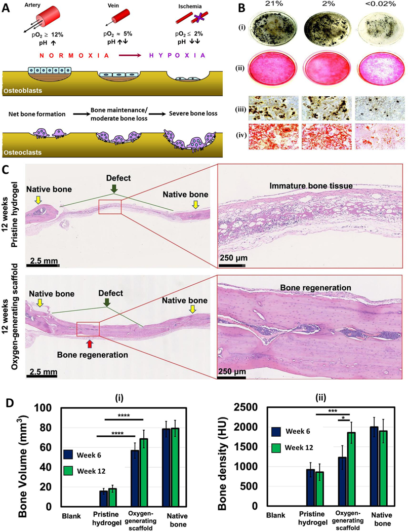
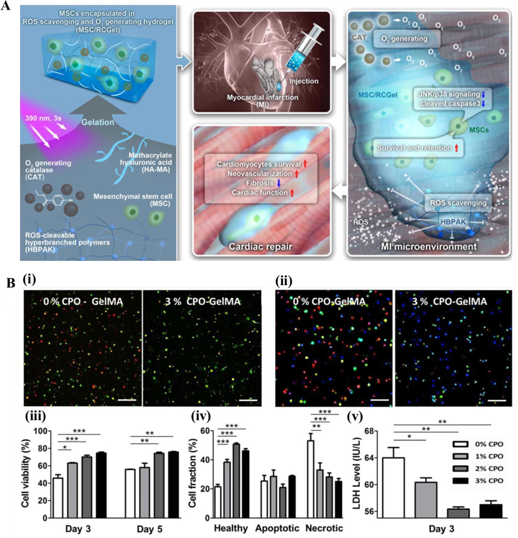
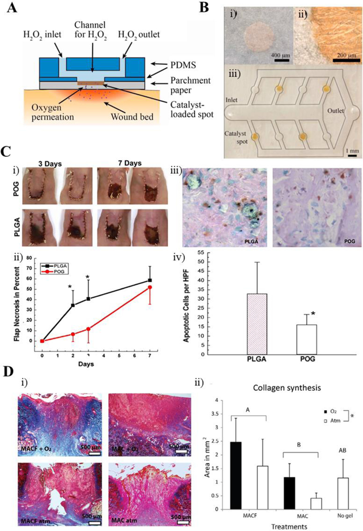
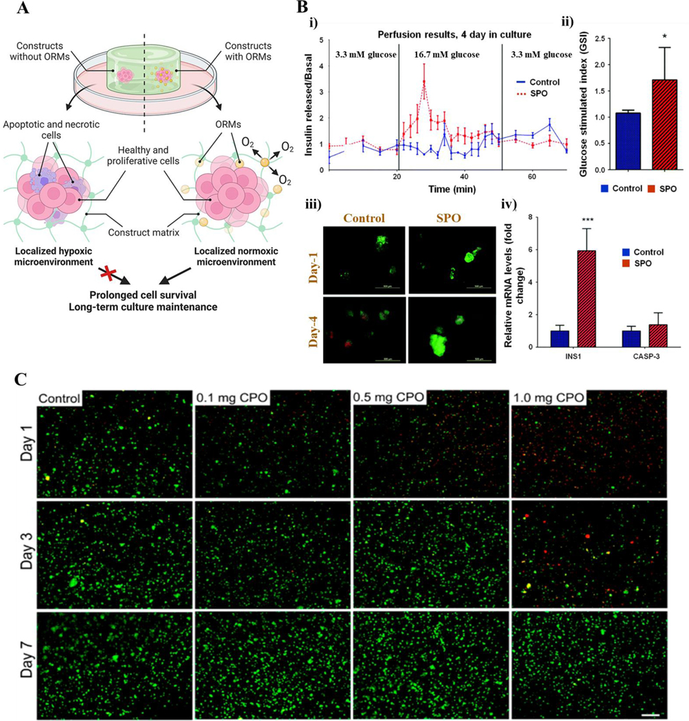
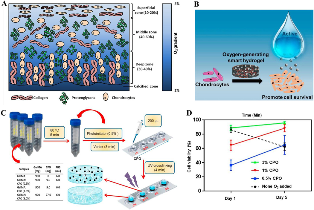
The limited availability of transplantable organs hinders the success of patient treatment through organ transplantation. In addition, there are challenges with immune rejection and the risk of disease transmission when receiving organs from other individuals. Tissue engineering aims to overcome these challenges by generating functional three-dimensional (3D) tissue constructs. When developing tissues or organs of a particular shape, structure, and size as determined by the specific needs of the therapeutic intervention, a tissue specific oxygen supply to all parts of the tissue construct is an utmost requirement. Moreover, the lack of a functional vasculature in engineered tissues decreases cell survival upon implantation in the body. Oxygen-generating materials can alleviate this challenge in engineered tissue constructs by providing oxygen in a sustained and controlled manner. Oxygen-generating materials can be incorporated into 3D scaffolds allowing the cells to receive and utilize oxygen efficiently. In this review, we present an overview of the use of oxygen-generating materials in various tissue engineering applications in an organ specific manner as well as their potential use in the clinic.


Figures

Figure 1.
Figure 1.
Oxygen-generating materials in bone tissue engineering. A) A scheme showing the effect of oxygen partial pressure in osteoblast and osteoclast activity during bone regeneration. B) Influence of oxygen supply on osteogenic cell survival, maintenance, and differentiation as well as subsequent bone regeneration (studied in terms of before blood vessel formation and after blood vessel formation). C) Mouse osteoblast differentiation following a 24-h exposure to ambient air (21% O2), hypoxia (2% O2), or anoxia (<0.02% O2). Representative low magnification images of (i) von Kossa and (ii) alizarin red stained cells indicated decreased bone nodule formation following less than 0.02% O2 exposure compared with 21 and 2% O2. High magnification images of (ii) von Kossa and (iv) alizarin red stained cells confirmed limited mineralized nodule formation by the anoxia-treated group. D) A scheme showing the fabrication of polycaprolactone (PCL)/CaO2-based oxygen-generating microparticles and oxygen-generating scaffold. E) Bone volume and (ii) Bone density measurements as well as F) H & E-stained histology images of healed bone indicate that the oxygen-generating scaffolds better support bone regeneration in comparison with the pristine scaffolds. Fig. A is reproduced from Ref. (82) with the permission of Springer-Nature, copyright 2013. Fig. B is reproduced from Ref. (83) with the creative common attribution license (CC-BY-0.4), copyright 2004. Fig. C, D are reproduced from Ref. (77) with the permission of Elsevier, copyright 2022.
Figure 2.
Figure 2.
Oxygen-generating materials in cardiac tissue engineering. A) General overview of cell-based therapies employed in cardiac tissue engineering and the importance of oxygen. The scheme shows that an oxygen-generating and ROS scavenging MSC/ hydrogel scaffold (MSC/RCGel), reduces fibrosis and promotes cell survival in an infarcted heart. B) Calcium peroxide (CPO) loaded oxygen generating GelMA hydrogels increase the survival of cardiac side population cells (CSPs) by preventing hypoxia induced necrosis. Confocal microphotographs of (i) live (green)/dead (red) and (ii) healthy (blue), apoptotic (green), and necrotic (red) CSPs cultured under hypoxic conditions in CPO loaded GelMA. Semiquantification of viable cells (iii) as well as (iv) healthy, apoptotic, and necrotic cells. (v) LDH activity of CSP cultured in CPO loaded GelMA hydrogels. Scale bar equals 50 micrometers. 297×150mm (300 × 300 DPI) Fig. A is reproduced from Ref. (30) with the permission of Elsevier, copyright 2022. Fig. B is reproduced from Ref. (67) with the permission of the American Chemical Society, copyright 2017.
Figure 3.
Figure 3.
Oxygen-generating materials in wound healing and regeneration. A) Schematic of a catalyzed oxygen-generating paper-based platform to be employed directly on the wound site. B) (i) Top and (ii) magnified views of the catalyst-loaded spots within the parchment paper-based platform as well as (iii) scheme showing the channels for hydrogen peroxide flow. C) Sodium percarbonate (SPO) laden PLGA films were implanted in mice models. Hematoxylin and eosin staining of skin flaps harvested at both 3 and 7 days. Delayed necrosis is seen in the SPO containing PLGA films (POG) through preservation of tissue architecture, hair follicles, epidermis height, and sebaceous glands compared to the non-oxygen-generating control films. D) Perfluorocarbon chains (MACF) based hydrogels were applied on dermal excisional wounds in rats. Representative Masson’s trichrome histology images for MACF and non-fluorinated methacrylamide chitosan (MAC) groups both with and without oxygen, as well as the no-gel control. New collagen synthesis is shown for each group, confirming the benefits of oxygen saturation of perfluorocarbon chains in the MACF group in wound healing. Fig. A, B are reproduced from Ref. (68) with permission from Elsevier, copyright 2014. Fig. C is reproduced from Ref. (69) with the permission of Elsevier, copyright 2007. Fig. D is reproduced from Ref. (70) with the permission of Elsevier, copyright 2016.
Figure 4.
Figure 4.
Oxygen-generating materials in soft tissue engineering. A) Schematic showing the role of oxygen-generating/releasing materials (ORMs) and their ability to enhance cell survival in soft tissue engineering applications. B) Incorporation of sodium percarbonate (SPO) on islet cell/PDMS substrate cultures showed greater cell proliferation and viability as seen through i) perfusion results, ii) glucose stimulated index, iii) representative live/dead fluorescent micrographs, and iv) profiling of INS1 and CASP-3 genes of cell cultures, when compared to the non-SPO control cultures. Myosin heavy chain and DAPI (blue) nuclei staining confirms the higher rates of cellular differentiation of SPO-containing cultures. C) Live (green)/dead (red) staining of C2C12 in muscle cells within the GelMA bioinks at different CaO2 concentrations crosslinked with UV light. This study found the optimal CaO2 concentration to be included into the scaffold was 0.5 mg for maintaining and enhancing cell viability and activity. Fig. A is reproduced from Ref. (25) with permission from Elsevier, copyright 2021. Fig. B is modified and reprinted from Ref. (85) with permission from the Royal Society of Chemistry (RSC), copyright 2017. Fig. C is reproduced from Ref. (73) under the creative common attribution license (CC-BY-0.4), copyright 2017.
Figure 5.
Figure 5.
Oxygen-generating materials in cartilage tissue engineering. A) Schematic showing the oxygen-gradient in various regions of articular cartilage. B) Schematic detailing how oxygen-generating hydrogels supplement chondrocytes with oxygen to promote cell viability. C) Representation of the fabrication process of the oxygen-releasing “smart” hydrogels at various CPO (CaO2) concentrations. GelMA was mixed with CaO2 and the photoinitiator; the mixture was subsequently UV-crosslinked to form the constructs. D) Viability of chondrocytes in GelMA hydrogels from day 1 and day 5 under oxygen-free conditions. Fig. A is reproduced from Ref. (112) under the creative common attribution license (CC-BY-0.4), copyright 2019. Fig. B, C, D are reproduced from Ref. (110) with permission from Elsevier, copyright 2020.

Similar articles

Cited by

References

    1. Morrison E, Suvarnapathaki S, Blake L, Camci-Unal G. Unconventional biomaterials for cardiovascular tissue engineering. Current Opinion in Biomedical Engineering. 2021;17:100263.
    1. Gómez MP, Pérez B, Manyalich M. International Registry in Organ Donation and Transplantation—2013. Transplantation Proceedings. 2014;46(4):1044–8. - PubMed
    1. Sadeghi F, Ramezani M, Beigee FS, Shadnia S, Moghaddam HH, Zamani N, et al. Organ Procurement From Poisoned Patients: A 14-Year Survey in 2 Academic Centers. Exp Clin Transplant. 2021. - PubMed
    1. Langer R, Vacanti Joseph P. Tissue Engineering. Science. 1993;260(5110):920–6. - PubMed
    1. Bioengineering NioBIa. Tissue Engineering and Regenerative Medicine. In: Services USDoHaH, editor. https://wwwnibibnihgov/sites/default/files/2020-06/Tissue_Engineering_Fa.... Bethesda, MD: National Institutes of Health; 2019. p. 1–2.