. 2023 Feb 28;97(2):e0144422.
doi: 10.1128/jvi.01444-22. Epub 2023 Jan 23.

A Zinc Finger Motif in the P1 N Terminus, Highly Conserved in a Subset of Potyviruses, Is Associated with the Host Range and Fitness of Telosma Mosaic Virus

Affiliations

A Zinc Finger Motif in the P1 N Terminus, Highly Conserved in a Subset of Potyviruses, Is Associated with the Host Range and Fitness of Telosma Mosaic Virus

Bei Gou et al. J Virol. .

Abstract

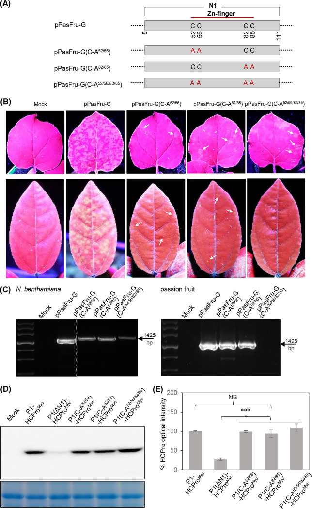
P1 is the first protein translated from the genomes of most viruses in the family Potyviridae, and it contains a C-terminal serine-protease domain that cis-cleaves the junction between P1 and HCPro in most cases. Intriguingly, P1 is the most divergent among all mature viral factors, and its roles during viral infection are still far from understood. In this study, we found that telosma mosaic virus (TelMV, genus Potyvirus) in passion fruit, unlike TelMV isolates present in other hosts, has two stretches at the P1 N terminus, named N1 and N2, with N1 harboring a Zn finger motif. Further analysis revealed that at least 14 different potyviruses, mostly belonging to the bean common mosaic virus subgroup, encode a domain equivalent to N1. Using the newly developed TelMV infectious cDNA clones from passion fruit, we demonstrated that N1, but not N2, is crucial for viral infection in both Nicotiana benthamiana and passion fruit. The regulatory effects of N1 domain on P1 cis cleavage, as well as the accumulation and RNA silencing suppression (RSS) activity of its cognate HCPro, were comprehensively investigated. We found that N1 deletion decreases HCPro abundance at the posttranslational level, likely by impairing P1 cis cleavage, thus reducing HCPro-mediated RSS activity. Remarkably, disruption of the Zn finger motif in N1 did not impair P1 cis cleavage and HCPro accumulation but severely debilitated TelMV fitness. Therefore, our results suggest that the Zn finger motif in P1s plays a critical role in viral infection that is independent of P1 protease activity and self-release, as well as HCPro accumulation and silencing suppression. IMPORTANCE Viruses belonging to the family Potyviridae represent the largest group of plant-infecting RNA viruses, including a variety of agriculturally and economically important viral pathogens. Like all picorna-like viruses, potyvirids employ polyprotein processing as the gene expression strategy. P1, the first protein translated from most potyvirid genomes, is the most variable viral factor and has attracted great scientific interest. Here, we defined a Zn finger motif-encompassing domain (N1) at the N terminus of P1 among diverse potyviruses phylogenetically related to bean common mosaic virus. Using TelMV as a model virus, we demonstrated that the N1 domain is key for viral infection, as it is involved both in regulating the abundance of its cognate HCPro and in an as-yet-undefined key function unrelated to protease processing and RNA silencing suppression. These results advance our knowledge of the hypervariable potyvirid P1s and highlight the importance for infection of a previously unstudied Zn finger domain at the P1 N terminus.

Keywords: HCPro; P1; Potyviridae; Potyvirus; RNA silencing suppression; Zn finger motif; host fitness; telosma mosaic virus.


Conflict of interest statement

The authors declare no conflict of interest.

Figures

FIG 1
FIG 1
Multiple alignment of amino acid sequences of TelMV P1s. The serine protease domain and amino-terminal stretches N1 and N2 in P1 are shown. A triad of amino acid residues (His-Gly-Ser) as putative catalytic activity sites are indicated by asterisks. These sequences were retrieved from NCBI GenBank database, and their accession numbers are MG944249 (TelMV-PasFru), MK340754 (TelMV-Fuzhou), MK340755 (TelMV-Wuyishan), and NC_009742 (TelMV-Hanoi). The genome of TelMV-Patchouli from patchouli was reconstructed from RNA-seq data in this study (Data Set S1).
FIG 2
FIG 2
Multiple amino acid sequence alignment of N1-containing potyviral P1s. Identical residues are marked with asterisks, and the catalytic triad residues His-Glu/Asp-Ser in the serine protease domain are indicated by stars. Alignment quality is shown as a bar graph below the alignment. The accession numbers of these sequences in GenBank database are MH286883 for passion fruit Vietnam virus (PVNV), KC845322 for soybean mosaic virus (SMV), MK217416 for watermelon mosaic virus (WMV), MK069986 for bean common mosaic virus (BCMV), LC377302 for East Asian Passiflora distortion virus (EAPDV), KT724930 for East Asian Passiflora virus (EAPV), MG197985 for papaya leaf distortion mosaic virus (PLDMV), LC228573 for gomphocarpus mosaic virus (GoMV), MK241979 for dendrobium chlorotic mosaic virus (DeCMV), EU410442 for Algerian watermelon mosaic virus (AWMV), MN124782 for cowpea aphid-borne mosaic virus (CABMV), MH376747 for pleione flower breaking virus (PlFBV), and MN549985 for paris virus 1 (ParV1).
FIG 3
FIG 3
Phylogenetic relationships of 136 virus species within the genus Potyvirus. The amino acid sequence alignment of complete polyprotein of these virus species was retrieved from the ICTV webpage (https://ictv.global/report/chapter/potyviridae/potyviridae/resources) (5). The pairwise unrooted maximum-likelihood tree was generated using PhyML v.3.0 (93) and visualized using iTOL (https://itol.embl.de/). The numbers at the branch nodes indicate bootstrap support (100 replicates), and values below 70% are not shown. The bar represents 0.5 substitution per site. The subgroups represented by corresponding viruses are defined with reference to a recent publication (5). A subset of potyviruses having the Zn finger-encompassing N1 domain are indicated with red dots.
FIG 4
FIG 4
Development of infections cDNA clones of TelMV-PasFru. (A) Diagram representing the genomic organization of TelMV-PasFru. “VPg” and “(A)n” at the 5′ and 3′ ends, respectively, denote the genome-linked viral protein VPg and poly(A). The 5′ and 3′ untranslated regions are indicated by two short horizontal lines. The large rectangle and short bar indicate the corresponding long and small ORFs. (B) Schematic representation of the full-length cDNA clone of TelMV-PasFru (pPasFru). The full-length cDNA of TelMV-PasFru was placed between the 35S promoter and Nos terminator in a modified T-DNA vector. Nucleotides belonging to pCB301 backbone and virus are in lowercase and uppercase, respectively. The MluI site, downstream of the poly(A) tail, is shown in italic type. (C) Schematic representation of pPasFru and pPasFru-G. (D) Infectivity test of pPasFru in N. benthamiana. Representative photographs of infiltrated plants were taken at 15 dpi. The leaves indicated by the box are enlarged in the lower images. Mock, empty vector control. (E) Transmission electron micrograph of TelMV particle. Bar, 200 nm. (F) RT-PCR detection of TelMV. The systemic leaves of N. benthamiana plants at 15 dpi (top) and passion fruit seedlings at 20 dpri (bottom) were sampled. (G) Infectivity test of pPasFru-G in N. benthamiana. Representative photographs were taken under a UV lamp at 10 dpi. Mock, empty vector control. (H) Western blot detection of GFP in upper noninoculated leaves of N. benthamiana (at 10 dpi) and passion fruit (at 20 dpri). (I and J) Infectivity test of virus progeny derived from pPasFru and pPasFru-G in passion fruit.
FIG 5
FIG 5
Infectivity test of N1 and N2 deletion TelMV clones in N. benthamiana and passion fruit. (A) Schematic diagrams of pPasFru-G and its truncated mutants. The ΔN1 and ΔN2 deletions are represented with labeled black bars. (B) Infectivity test of the indicated virus clones in N. benthamiana and passion fruit. Representative photographs were taken under a UV lamp at 12 dpi for N. benthamiana and at 18 dpri for passion fruit. (C) RT-PCR detection of virus progeny derived from the indicated virus clones. The upper noninoculated leaves of N. benthamiana (at 12 dpi) and passion fruit (at 18 dpri) were sampled for the assay. (D) Immunoblot detection of GFP in upper noninoculated leaves of N. benthamiana (at 12 dpi) and passion fruit (at 18 dpri).
FIG 6
FIG 6
Effects of N1 deletion in TelMV clone on viral intercellular movement and replication. (A) Time course observation of viral intercellular movement for the indicated virus clones. The viral intercellular movement from single primarily infected cells was monitored at 48, 60, 72, and 96 hpi. Bars, 100 nm. (B) Statistic analysis of spreading area of viral infection foci at 72 hpi. Error bars denote standard errors for at least 25 infection foci for each clone from three independent experiments. *, 0.01 < P < 0.05. (C and D) Effects of N1 deletion in TelMV clone on viral genomic RNA and protein accumulations. Viral genomic RNA accumulations were determined by real-time RT-qPCR (C), and GFP accumulation by immunoblot assay (D). Error bars denote the standard errors from three biological replicates. *, 0.01 < P < 0.05; **, 0.001 < P < 0.01.
FIG 7
FIG 7
Effects of N1 deletion in P1 on the abundance of its cognate HCPro. (A) Schematic diagrams of P1-HCProMyc, P1ΔN1-HCProMyc, P1-MycHCPro-GFP, and P1ΔN1-MycHCPro-GFP. The black bar labeled “ΔN1” represents N1 deletion. Red arrows denote cis cleavage sites of P1 or HCPro. (B) Immunoblot detection of HCProMyc abundance in N. benthamiana leaves. (C) Quantitative analysis of HCProMyc signals in panel B at 2 dpi. The signal intensity values are presented as means and standard deviations (SD) (n = 3). The average value for P1-HCProMyc was designated 100% to normalize the data. *, 0.01 < P < 0.05. (D) Real-time RT-qPCR assay of the mRNA transcripts from N. benthamiana leaves infiltrated with pCaM-P1-HCProMyc and pCaM-P1ΔN1-HCProMyc. Agrobacterial cultures harboring relevant plasmids were adjusted to an OD600 of 0.3 and infiltrated into N. benthamiana leaves. The infiltrated leaves were sampled at 48 hpi for real-time RT-qPCR assay. Error bars denote the standard errors from three biological replicates. NS, no significant difference. (E) Immunoblot detection of MycHCPro accumulation levels. Agrobacterial cultures harboring relevant plasmids were adjusted to an OD600 of 0.3 for infiltration into N. benthamiana leaves, and the treated leaves were sampled at 48 hpi for Western blot assay. (F) Quantitative analysis of MycHCPro signals in panel E. The signal intensity values are presented as means and SD (n = 3). The average values for P1-MycHCPro-GFP were designated 100% to normalize the data. *, 0.01 < P < 0.05. (G) Immunoblot detection of GFP accumulation levels. (H) Quantitative analysis of GFP signals in panel G. NS, no significant difference.
FIG 8
FIG 8
The N1 domain facilitates P1 cis cleavage and thus enhances the abundance of HCPro. (A) Schematic diagrams of P1-HCProMyc, P1ΔN1-HCProMyc, P1(S/A)-HCProMyc, P1ΔN1(S/A)-HCProMyc, P1(S/A-CS)-HCProMyc, and P1ΔN1(S/A-CS)-HCProMyc. (B) Immunoblot analysis of HCProMyc abundance from N. benthamiana leaves coexpressing TelMV NIa and the indicated proteins. Mock, empty vector control. Quantitative analysis of HCProMyc signals is shown (bottom). The signal intensity values are presented as means and SD (n = 3). The average value for NIa+P1-HCProMyc was designated 100% to normalize the data. **, 0.001 < P < 0.01. (C) Real-time RT-qPCR assay of mRNA transcripts of the indicated proteins. The coinfiltrated N. benthamiana leaves were sampled at 48 hpi for the assay. Error bars denote standard errors from three biological replicates. NS, no significant difference.
FIG 9
FIG 9
The N1 domain in P1 modulates the abundance of HCPro and its RSS activity in the presence of other viral proteins (P3 to CP). (A) Schematic diagrams of four T-DNA constructs (pP3-CP-GFP, pP3-CP-GFP//P1-HCProMyc, pP3-CP-GFP//P1ΔN1-HCProMyc, and pP3-CP-GFP//HCProMyc). (B) Expression analysis of P3 to CP in N. benthamiana leaves. The mRNA transcripts of P3 to CP were detected by RT-PCR. The intracellular GFP signals were monitored by fluorescence microscopy. GFP was translated along with the truncated viral genome and released from polyprotein by proteolytic cleavage of NIa-Pro in trans. Lanes 1 and 2, empty vector control; lanes 3 and 4, pP3-CP-GFP. (C) Immunoblot analysis of HCProMyc accumulation in N. benthamiana leaves infiltrated with the indicated plasmids. (D) Quantitative analysis of HCProMyc signals in panel C. The signal intensity values are presented as means and SD (n = 3). The average value for pP3-CP-GFP//P1ΔN1-HCProMyc was designated 100% to normalize the data. *, 0.01 < P < 0.05. (E) RSS activity test of P1-HCProMyc, P1ΔN1-HCProMyc, and HCProMyc in the presence of viral proteins P3 to CP. Representative photographs of coinfiltrated N. benthamiana leaves were taken at 2 dpi. The patch design for coinfiltration was as follows: a GFP-expressing plasmid, together with a candidate protein-expressing construct (indicated by 3), P19-expressing construct (positive control, indicated by 1), or empty vector pCaMterX (negative control, indicated by 2). (F) Immunoblot detection of GFP accumulation from coinfiltrated leaf patches at 2 dpi. Coomassie blue staining of the large subunit of RubisCO was used as the loading control. Quantitative analysis of GFP signals is shown (bottom). The signal intensity values are presented as means and SD (n = 3). The average value for P3-CP//P1-HCProMyc was designated 100% to normalize the data. *, 0.01 < P < 0.05. (G) Northern blot analysis of GFP mRNA accumulation in coinfiltrated leaf patches at 2 dpi. GelRed staining of rRNA served as the loading control. (H) Real-time RT-qPCR assay of GFP mRNA transcripts from coinfiltrated N. benthamiana leaves. The samples were collected at 48 hpi for the assay. Error bars denote the standard errors from three biological replicates. *, 0.01 < P < 0.05; **, 0.001 < P < 0.01.
FIG 10
FIG 10
Disruption of Zn finger motif in N1 debilitates viral compatible infection but does not affect P1 cis cleavage and HCPro accumulation. (A) Schematic diagrams of pPasFru-G and its mutated versions. (B) Infectivity test of the indicated virus clones in N. benthamiana and passion fruit. Representative photographs of upper noninoculated leaves were taken at 15 dpi for N. benthamiana and 18 dpri for passion fruit. The scattered weak fluorescence spots are indicated by arrows. (C) RT-PCR detection of virus progeny derived from the indicated virus clones. The upper noninoculated leaves of N. benthamiana at 15 dpi (left) and passion fruit at 18 dpri (right) were sampled. (D) Immunoblot detection of HCProMyc from N. benthamiana leaves inoculated with the relevant plasmids. (E) Quantitative analysis of HCProMyc signals in panel D. The signal intensity values are presented as means and SD (n = 3). The average value for P1-HCProMyc was designated 100% to normalize the data. ***, P < 0.001; NS, no significant difference.
FIG 11
FIG 11
Working model depicting the involvement of the N1 domain of TelMV P1 in viral host fitness.

References

    1. Revers F, García JA. 2015. Molecular biology of potyviruses. Adv Virus Res 92:101–199. 10.1016/bs.aivir.2014.11.006. - DOI - PubMed
    1. Cui H, Wang A. 2019. The biological impact of the hypervariable N-terminal region of potyviral genomes. Annu Rev Virol 6:255–274. 10.1146/annurev-virology-092818-015843. - DOI - PubMed
    1. Yang X, Li Y, Wang A. 2021. Research advances in potyviruses: from the laboratory bench to the field. Annu Rev Phytopathol 59:1–29. 10.1146/annurev-phyto-020620-114550. - DOI - PubMed
    1. Wylie SJ, Adams M, Chalam C, Kreuze J, López-Moya JJ, Ohshima K, Praveen S, Rabenstein F, Stenger D, Wang A, Zerbini FM, ICTV Report Consortium . 2017. ICTV virus taxonomy profile: Potyviridae. J Gen Virol 98:352–354. 10.1099/jgv.0.000740. - DOI - PMC - PubMed
    1. Inoue-Nagata AK, Jordan R, Kreuze J, Li F, López-Moya JJ, Mäkinen K, Ohshima K, Wylie SJ, ICTV Report Consortium . 2022. ICTV virus taxonomy profile: Potyviridae 2022. J Gen Virol 103:e001738. 10.1099/jgv.0.001738. - DOI - PubMed

Publication types

Supplementary concepts

LinkOut - more resources