. 2023 Feb 28;97(2):e0171622.
doi: 10.1128/jvi.01716-22. Epub 2023 Jan 23.

Minimal Antigenic Evolution after a Decade of Norovirus GII.4 Sydney_2012 Circulation in Humans

Affiliations

Minimal Antigenic Evolution after a Decade of Norovirus GII.4 Sydney_2012 Circulation in Humans

Gabriel I Parra et al. J Virol. .

Abstract

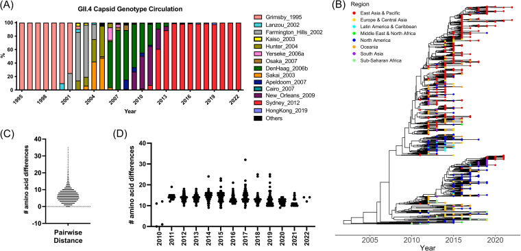
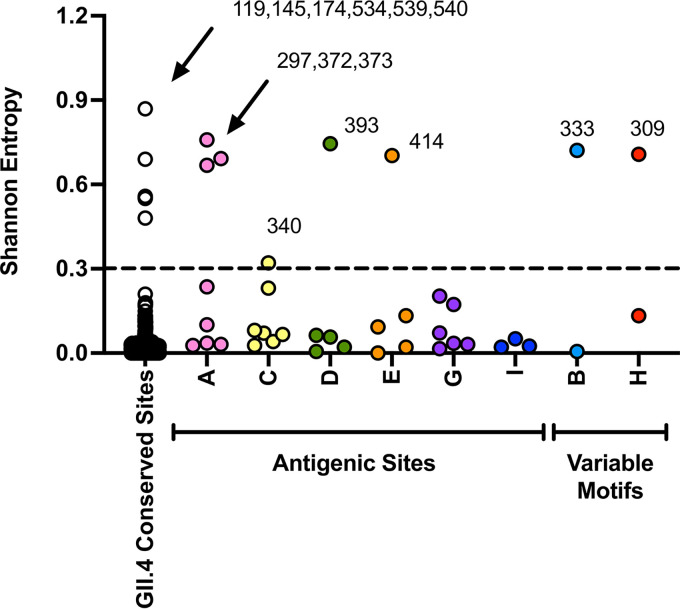
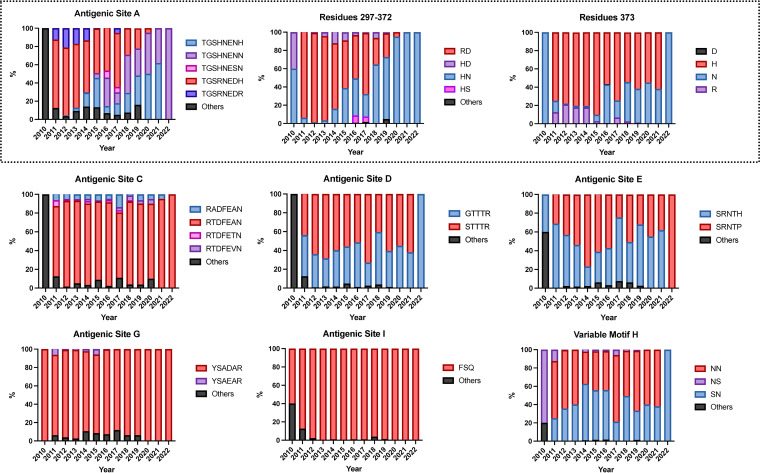
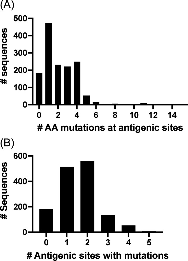
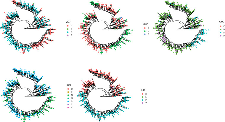
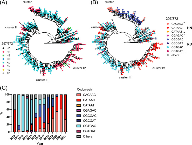
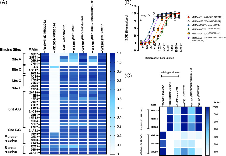
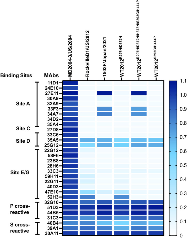
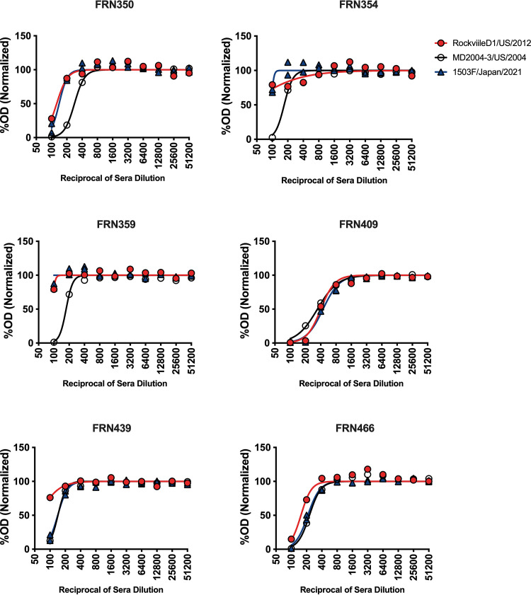
Norovirus is a major human pathogen that can cause severe gastroenteritis in vulnerable populations. The extensive viral diversity presented by human noroviruses constitutes a major roadblock for the development of effective vaccines. In addition to the large number of genotypes, antigenically distinct variants of GII.4 noroviruses have chronologically emerged over the last 3 decades. The last variant to emerge, Sydney_2012, has been circulating at high incidence worldwide for over a decade. We analyzed 1449 capsid sequences from GII.4 Sydney_2012 viruses to determine genetic changes indicative of antigenic diversification. Phylogenetic analyses show that Sydney_2012 viruses scattered within the tree topology with no single cluster dominating during a given year or geographical location. Fourteen residues presented high variability, 7 of which mapped to 4 antigenic sites. Notably, ~52% of viruses presented mutations at 2 or more antigenic sites. Mutational patterns showed that residues 297 and 372, which map to antigenic site A, changed over time. Virus-like particles (VLPs) developed from wild-type Sydney_2012 viruses and engineered to display all mutations detected at antigenic sites were tested against polyclonal sera and monoclonal antibodies raised against Sydney_2012 and Farmington_Hills_2002 VLPs. Minimal changes in reactivity were detected with polyclonal sera and only 4 MAbs lost binding, with all mapping to antigenic site A. Notably, reversion of residues from Sydney_2012 reconstituted epitopes from ancestral GII.4 variants. Overall, this study demonstrates that, despite circulating for over a decade, Sydney_2012 viruses present minimal antigenic diversification and provides novel insights on the diversification of GII.4 noroviruses that could inform vaccine design. IMPORTANCE GII.4 noroviruses are the major cause of acute gastroenteritis in all age groups. This predominance has been attributed to the continued emergence of phylogenetically discrete variants that escape immune responses to previous infections. The last GII.4 variant to emerge, Sydney_2012, has been circulating at high incidence for over a decade, raising the question of whether this variant is undergoing antigenic diversification without presenting a major distinction at the phylogenetic level. Sequence analyses that include >1400 capsid sequences from GII.4 Sydney_2012 showed changes in 4 out of the 6 major antigenic sites. Notably, while changes were detected in one of the most immunodominant sites over time, these resulted in minimal changes in the antigenic profile of these viruses. This study provides new insights on the mechanism governing the antigenic diversification of GII.4 norovirus that could help in the development of cross-protective vaccines to human noroviruses.

Keywords: antigenic variation; calicivirus; gastroenteritis; norovirus; phylogenetic analysis.


Conflict of interest statement

The authors declare no conflict of interest.

Figures

FIG 1
FIG 1
Evolution of the major capsid protein (VP1) from GII.4 Sydney_2012 noroviruses. (A) GII.4 variant yearly distribution tabulated using all sequences available in public databases (11). The last variant to emerge, GII.4 Sydney_2012 variant, has been circulating for over a decade. (B) Time-scaled phylogenetic tree of GII.4 Sydney_2012 noroviruses showing the circulation of different clusters every given year. The phylogenetic tree was calculated using 1,449 nucleotide sequences encoding the VP1 of Sydney_2012 noroviruses and the Markov chain Monte Carlo (MCMC) method, as implemented in BEAST v1.10.4 (41). (C) Amino acid pairwise differences among all Sydney_2012 viruses calculated using the same data set used for phylogenetic tree reconstruction. (D) Amino acid pairwise differences of all Sydney_2012 viruses in each year compared to the earliest virus detected in 2010. Intra-variant diversification reveals an overall variation of 2 to 11 (10th to 90th percentile; range 1 to 35) residues.
FIG 2
FIG 2
Conservation analysis of the major capsid protein (VP1) from GII.4 Sydney_2012 noroviruses. Shannon entropy was used to calculate the amino acid variation for each site in the VP1. Analysis was carried out using 1,449 complete (or nearly complete) sequences of Sydney_2012 noroviruses and the Shannon Entropy-One tool, as implemented in Los Alamos National Laboratory. Residues were grouped on those mapping outside antigenic sites (non-antigenic sites) and those mapping on antigenic sites (columns A to E, G to I). Residues presenting entropy values ≥0.3 are considered as variables (13). Variable motifs are predicted to be relevant to virus diversification, but not yet experimentally demonstrated to be an antigenic site.
FIG 3
FIG 3
Temporal amino acid patterns on the major antigenic sites from GII.4 Sydney_2012 noroviruses. Colors of the bars correspond to the most frequent sequence patterns presented at any given antigenic site. Amino acid changes occurring on the 3 (297, 372, and 373) variable residues from antigenic site A are shown separately. Analysis was carried out using 1,449 complete (or nearly complete) sequences of Sydney_2012 noroviruses and scripts implemented in R (13, 38).
FIG 4
FIG 4
Quantification of amino acid mutations on the major antigenic sites from GII.4 Sydney_2012 noroviruses. (A) Distribution of viruses by the number of mutations presented at antigenic sites as compared with the consensus sequence of Sydney_2012 variant. (B) Distribution of viruses by the number of antigenic sites presenting any one mutation as compared with the consensus sequence of Sydney_2012 variant. The consensus sequence for the Sydney_2012 variant was calculated from 1,449 complete (or nearly complete) sequences of Sydney_2012 noroviruses and the Entropy tool as described in Materials and Methods.
FIG 5
FIG 5
Mutations on antigenic sites are associated to specific phylogenetic clusters. The tips of the time-scaled phylogenetic tree were color-coded based on the residue presented by every viral sequence. The phylogenetic trees were calculated as indicated in Fig. 1 and the Materials and Methods section.
FIG 6
FIG 6
Amino acid mutational pattern from antigenic site A from GII.4 Sydney_2012 variant is associated to differential genetic codon usage. (A) Amino acid distribution in the phylogenetic trees shows evolutionary convergence in residues 297 and 372 from major capsid protein (VP1) of GII.4 Sydney_2012 noroviruses. (B) Phylogenetic tree color-coded based on the codon presented for the codon pair encoding residues 297 and 372. (C) Temporal genetic diversification of the codon pair encoding amino acids 297 and 372. Colors of the bars correspond to the most frequent sequence patterns presented at those 2 codon positions. The analyses were done using 1,449 complete (or nearly complete) sequences of Sydney_2012 noroviruses. The phylogenetic trees were calculated as indicated in Fig. 1 and the Materials and Methods section. Clusters were arbitrarily assigned, in order to facilitate description, to monophyletic branching of strains with similar codon pair usage.
FIG 7
FIG 7
GII.4 Sydney_2012 noroviruses present minimal intra-variant antigenic variation. (A) A monoclonal antibody (MAb) library developed against a wild-type GII.4 Sydney_2012 virus (RockvilleD1/US/2012) indicates binding lost only by a subset of MAbs targeting antigenic site A. The heatmap indicates MAb-VLP binding as detected by ELISA, and represents the normalized OD405nm values. The columns denote wild-type and mutant VLPs. The OD405 nm were normalized to the cross-reactive MAb 30A11 (1.0) and the negative control (0). (B) Polyclonal sera responses from animals immunized with RockvilleD1/US/2012 VLPs. Normalized curves are shown from 1 mouse polyclonal serum as example. (C) The heatmap indicates the EC50 value from HBGA-blocking titers from the polyclonal sera from animals immunized with RoclvilleD1/US/2012 and MD2004-3/US/2004 (a wild-type Farmington_Hills_2002 virus) VLPs. The columns denote wild-type and mutant VLPs, and the rows show individual polyclonal sera.
FIG 8
FIG 8
GII.4 Sydney_2012 noroviruses present epitopes from ancestral GII.4 variants. A monoclonal antibody (MAb) library developed against a wild-type GII.4 Farmington_Hills_2002 virus (MD2004-3/US/2004) indicates reactivity of MAbs, mapping to antigenic site A, against newer Sydney_2012 viruses. The columns denote wild-type and mutant VLPs. The background of the mutant VLPs is that from the wild-type RockvilleD1/US/2012 virus. The OD405 nm were normalized to the cross-reactive MAb 30A11 (1.0) and the negative control (0).
FIG 9
FIG 9
Sera from individuals challenged with a Farmington_Hills_2002 virus present HBGA-blocking activity against modern Sydney_2012 VLPs. Normalized curves are shown from sera from 6 individuals challenged with the 031693/US/2003 virus (22). Two individuals, FRN439 and FRN466, presented blocking titers that indicate closer antigenicity between the modern Sydney_2012 virus (1503F/Japan/2021) and the Farmington_Hills_2002 virus (MD2004-3/US/2004). The 1503F/Japan/2021 virus presents an antigenic site A that resembles those from Farmington_Hills_2002 virus (Table 1).

References

    1. Ford-Siltz LA, Tohma K, Parra GI. 2021. Understanding the relationship between norovirus diversity and immunity. Gut Microbes 13:1–13. 10.1080/19490976.2021.1900994. - DOI - PMC - PubMed
    1. Ford-Siltz LA, Wales S, Tohma K, Gao Y, Parra GI. 2020. Genotype-specific neutralization of norovirus is mediated by antibodies against the protruding domain of the major capsid protein. J Infect Dis 225:1205–1214. 10.1093/infdis/jiaa116. - DOI - PubMed
    1. Prasad BV, Hardy ME, Dokland T, Bella J, Rossmann MG, Estes MK. 1999. X-ray crystallographic structure of the Norwalk virus capsid. Science 286:287–290. 10.1126/science.286.5438.287. - DOI - PubMed
    1. Marionneau S, Ruvoen N, Le Moullac-Vaidye B, Clement M, Cailleau-Thomas A, Ruiz-Palacois G, Huang P, Jiang X, Le Pendu J. 2002. Norwalk virus binds to histo-blood group antigens present on gastroduodenal epithelial cells of secretor individuals. Gastroenterology 122:1967–1977. 10.1053/gast.2002.33661. - DOI - PMC - PubMed
    1. Lindesmith L, Moe C, Marionneau S, Ruvoen N, Jiang X, Lindblad L, Stewart P, LePendu J, Baric R. 2003. Human susceptibility and resistance to Norwalk virus infection. Nat Med 9:548–553. 10.1038/nm860. - DOI - PubMed

Publication types