Review
. 2023 Apr;20(2):177-198.
doi: 10.1007/s13770-022-00510-z. Epub 2023 Jan 23.

Silica-Based Advanced Nanoparticles For Treating Ischemic Disease

Affiliations
Review

Silica-Based Advanced Nanoparticles For Treating Ischemic Disease

Dauletkerey Kudaibergen et al. Tissue Eng Regen Med. 2023 Apr.

Abstract

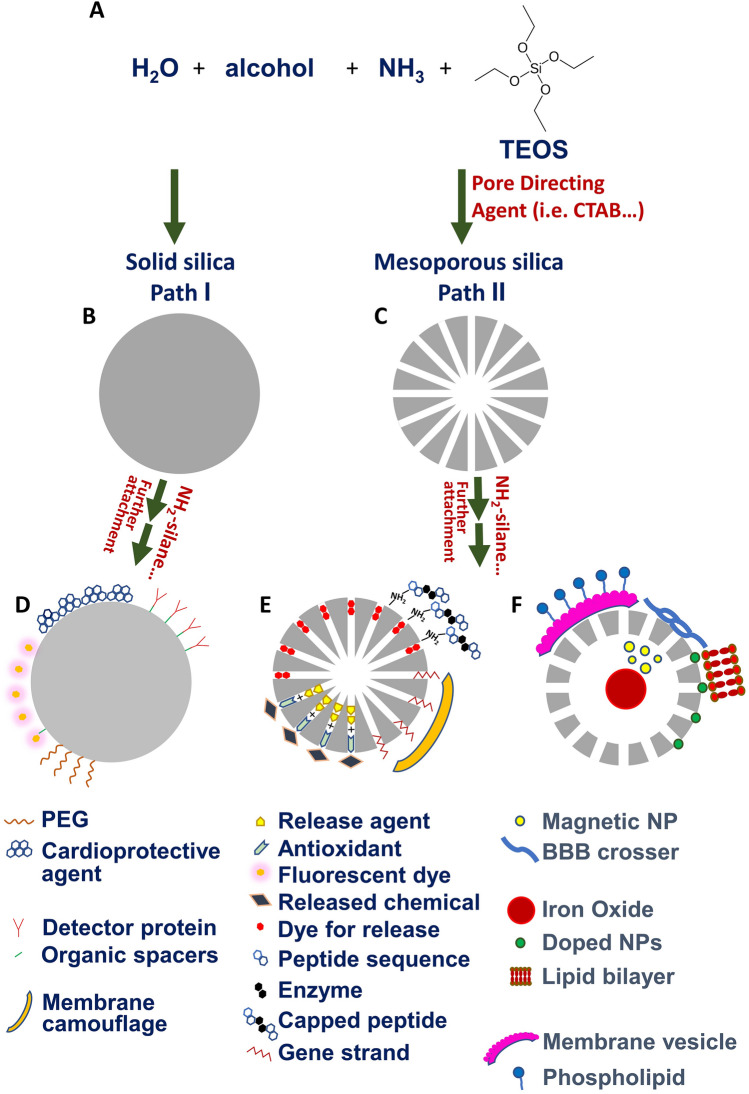
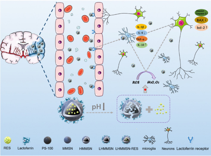
Recently, various attempts have been made to apply diverse types of nanoparticles in biotechnology. Silica nanoparticles (SNPs) have been highlighted and studied for their selective accumulation in diseased parts, strong physical and chemical stability, and low cytotoxicity. SNPs, in particular, are very suitable for use in drug delivery and bioimaging, and have been sought as a treatment for ischemic diseases. In addition, mesoporous silica nanoparticles have been confirmed to efficiently deliver various types of drugs owing to their porous structure. Moreover, there have been innovative attempts to treat ischemic diseases using SNPs, which utilize the effects of Si ions on cells to improve cell viability, migration enhancement, and phenotype modulation. Recently, external stimulus-responsive treatments that control the movement of magnetic SNPs using external magnetic fields have been studied. This review addresses several original attempts to treat ischemic diseases using SNPs, including particle synthesis methods, and presents perspectives on future research directions.

Keywords: Bioimaging; Drug delivery system; Ischemic disease treatment; Silica based magnetic nanoparticle; Silica nanoparticle.


Conflict of interest statement

The authors declare no conflict of interest.

Figures

Fig. 1
Fig. 1
A The fundamental components generally used for the synthesis of SNP. B Solid silica nanoparticle synthesized through Stober method, which includes silica precursor [i.e. TEOS], water, ammonia as a catalyst and alcohol as a solvent C Mesoporous silica nanoparticles synthesized through the addition of pore directing agents such as CTAB. Schematic diagram of D sSNPs and E mSNPs immobilized with various cargo molecules. F hollow mSNPs doped/loaded with metals and coated with different components
Fig. 2
Fig. 2
A TEM images of dye loaded mSNPs, which were capped with peptidic sequence exhibiting ordinary pore structure. Left part: as-prepared mSNPs (MCM-41). Middle part: mSNPs capped with S1-P1. Right part: mSNPs capped with S1-P2. Scale: 25 µm. B TEM images of MSN, cell-membrane camouflaged mesoporous silica nanoparticles (CM-MSN) and micro-RNA embedded CM-miR-MSNs. Scale: 500 nm. C TEM images of PS-100, hollow manganese doped mSNPs (HMMSN) and lactoferrin-functinalized hollow manganese doped mSNPs (LHMMSN). Scale: 100 nm. D TEM images of dense silica matrix coated with secondary mSNP shell (dSiO2@MSN), etched hollow mSNPs (hMSN), and MnCO-loaded, neutrophil membrane coated hollow mSNPs (MnCO@hMSN@NM-PS) negatively stained by phosphotungstic acid. Scale: 200 nm. A Reprinted with permission from Wiley, 2015 [30]. B Reprinted with permission from Elsevier, 2020 [31]. C Reprinted with permission from Elsevier, 2022 [33]. D Reprinted with permission from American Chemical Society, 2022 [35]
Fig. 3
Fig. 3
A Schematics delivery of tPA and SK through silica coated magnetic nanoparticles. B SEM image of structure and surface morphology corresponding to SiO2-MNP. C TEM image of polystyrene magnetite nano cluster (PMNC, left) and fluorescent magnetite nano clusters (FMNC, right). Scale: 100 nm. A, B Reprinted with permission from Elsevier, 2015 [36]. C Reprinted with permission from Elsevier, 2011 [37]
Fig. 4
Fig. 4
A Synthetic illustration demonstrated for TAP-SiO2@AuNPs for dual optical/computed tomography imaging B FE-TEM image of TAP-SiO2@AuNPs C Synthesis diagram of SIO nanostructures D Low magnification TEM image of SIO A, B Reprinted with permission from Elsevier, 2018 [39]. C, D Reprinted with permission from American Chemical Society, 2019 [40]
Fig. 5
Fig. 5
Drug delivery system using stem cell membrane-camouflaged exosome mimicking nanocomplex which contains miRNA in MSN. Detail mechanism of targeting ischemia area of cardiomyocytes. The CM-miRNA-MSN nanocomplex decomposes in cardiomyocytes and releases miRNA into cytoplasm inhibiting the translation of PTEN and PDCD4 to hamper apoptosis. Reprinted with permission from Elsevier, 2020 [31]
Fig. 6
Fig. 6
Action mechanism of LHMMSN in stroke treatment. Resveratrol loaded LHMMSN (LHMMSN-RES) degrade in low pH condition and release resveratrol. Detail mechanism of anti-inflammatory and neuroprotective effects were described. Reprinted with permission from Elsevier, 2022 [33]
Fig. 7
Fig. 7
A Illustration of NIRF and micro-computer tomography imaging mechanism using TAP-SiO2@AuNPs. B Illustration of treatment approach with SIO. Ultrasound and MRI contrast of hMSCs was increased by SIO. The NPs and an external magnet increase cell retention. Furthermore, the multifunctional silica-based NPs enhanced cell viability by sustained release of IGF. A Reprinted with permission from Elsevier, 2018 [39]. B Reprinted with permission from American Chemical Society, 2019 [40]
Fig. 8
Fig. 8
The expression of cardiac-specific genes and proteins in NRCMs which were cultured with each concentration of CS extracts for 7 days. A The expression of Myh6/Myh7, Tnnt2, Cacna1a and Gja1 genes in NRCMs cultured with the control medium and CS extracts with the dilution ratio from 1/1024 to 1/2. The immunofluorescent staining of cTnT protein B and Cx43 protein C) in NRCMs cultured with the control medium and CS extracts diluted at 1/256, 1/32 and 1/4 to confirm the expression amount. The second row shows the enlarged image (white square) and scale bars indicate 200 μm, 50 μm, 100 μm, 25 μm from the top row to bottom. *p < 0.05 and **p < 0.01 compared with blank compared with Blank, respectively (n = 4 each). A, B, C Reprinted with permission from American Chemical Society, 2019 [21]
Fig. 9
Fig. 9
The distribution of EPCs after being stimulated by external magnetic field. A These images show the distribution of magnetic nanoparticle labeled EPCs with or without an external magnetic field as external physical stimulus. B These images show the distribution of magnetic nanoparticle labeled EPCs with or without an external magnetic field as external physical stimulus at flowing condition. A, B Reprinted with permission from Wiley, 2019 [13]

Similar articles

Cited by

References

    1. Bobo D, Robinson KJ, Islam J, Thurecht KJ, Corrie SR. Nanoparticle-based medicines: a review of FDA-approved materials and clinical trials to date. Pharm Res. 2016;33:2373–2387. doi: 10.1007/s11095-016-1958-5. - DOI - PubMed
    1. Khalid K, Tan X, Mohd Zaid HF, Tao Y, Lye Chew C, Chu DT, et al. Advanced in developmental organic and inorganic nanomaterial: a review. Bioengineered. 2020;11:328–355. doi: 10.1080/21655979.2020.1736240. - DOI - PMC - PubMed
    1. Sur S, Rathore A, Dave V, Reddy KR, Chouhan RS, Sadhu V. Recent developments in functionalized polymer nanoparticles for efficient drug delivery system. Nano-Structures & Nano-Objects. 2019;20:100397. doi: 10.1016/j.nanoso.2019.100397. - DOI
    1. Desai N, Momin M, Khan T, Gharat S, Ningthoujam RS, Omri A. Metallic nanoparticles as drug delivery system for the treatment of cancer. Expert Opin Drug Deliv. 2021;18:1261–1290. doi: 10.1080/17425247.2021.1912008. - DOI - PubMed
    1. Bhattacharjee S, Rietjens IMCM, Singh MP, Atkins TM, Purkait TK, Zejing Xu, dSR, , et al. Cytotoxicity of surface-functionalized silicon and germanium nanoparticles: the dominant role of surface charges. Nanoscale. 2013;5:4870–4883. doi: 10.1039/c3nr34266b. - DOI - PMC - PubMed

Publication types

LinkOut - more resources