. 2023 Feb;54(2):354-363.
doi: 10.1161/STROKEAHA.122.041637. Epub 2023 Jan 23.

Worsened Stroke Outcome in a Model of Preeclampsia is Associated With Poor Collateral Flow and Oxidative Stress

Affiliations

Worsened Stroke Outcome in a Model of Preeclampsia is Associated With Poor Collateral Flow and Oxidative Stress

Marilyn J Cipolla et al. Stroke. 2023 Feb.

Abstract

Background: Preeclampsia increases the incidence of maternal stroke, a devastating condition that is on the rise. We investigated stroke outcome in a model of experimental preeclampsia with and without treatment with clinically relevant doses of magnesium sulfate (experimental preeclampsia+MgSO4) compared to normal late-pregnant and nonpregnant rats.

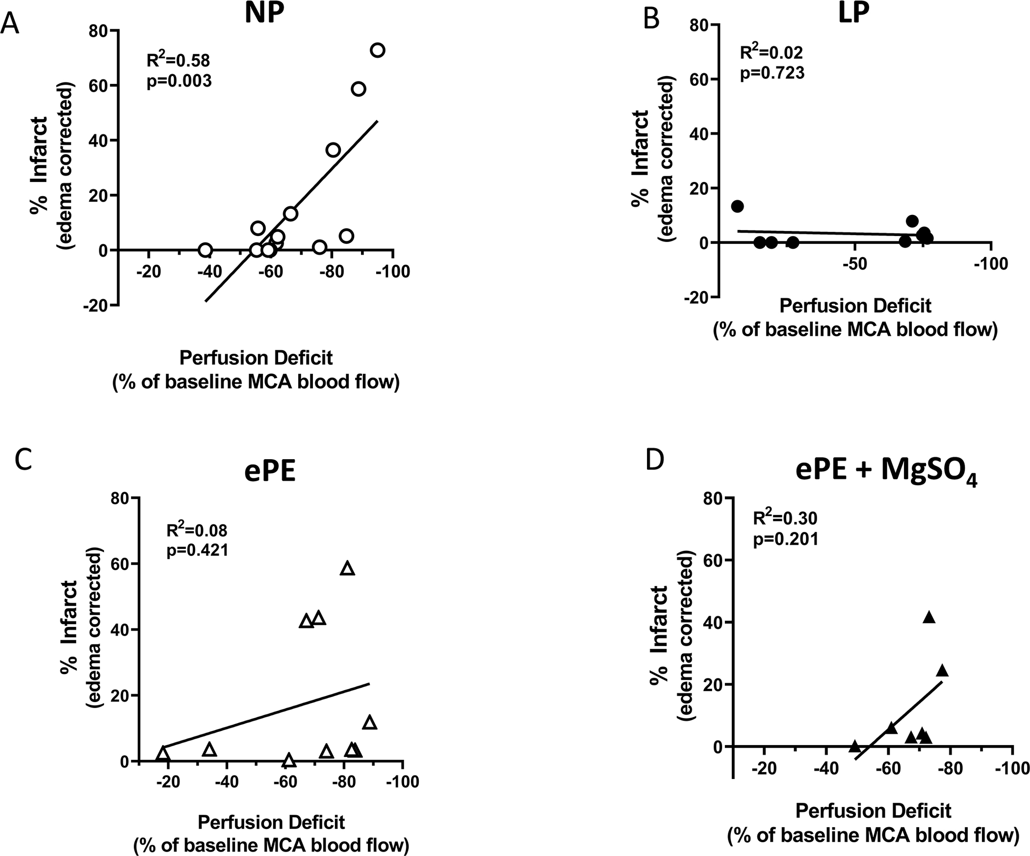
Methods: Transient middle cerebral artery occlusion was used to induce focal stroke for either 1.5 or 3 hours. Infarct volume and hemorrhagic transformation were determined as measures of stroke outcome. Changes in core middle cerebral artery and collateral flow were measured by dual laser Doppler. The relationship between middle cerebral artery perfusion deficit and infarction was used as a measure of ischemic tolerance. Oxidative stress and endothelial dysfunction were measured by 3-nitrotyrosine and 8-isoprostane, in brain and serum, respectively.

Results: Late-pregnant animals had robust collateral flow and greater ischemic tolerance of brain tissue, whereas experimental preeclampsia had greater infarction that was related to poor collateral flow, endothelial dysfunction, and oxidative stress. Importantly, pregnancy appeared preventative of hemorrhagic transformation as it occurred only in nonpregnant animals. MgSO4 did not provide benefit to experimental preeclampsia animals for infarction.

Conclusions: Stroke outcome was worse in a model of preeclampsia. As preeclampsia increases the risk of future stroke and cardiovascular disease, it is worth understanding the influence of preeclampsia on the material brain and factors that might potentiate injury both during the index pregnancy and years postpartum.

Keywords: brain; ischemia; preeclampsia; pregnancy; stroke.


Figures

Figure 1.
Figure 1.. Perfusion deficit and infarct volume after 1.5 hours of ischemia with 24 hours of reperfusion.
(A) Percent change in core MCA flow during filament occlusion calculated from baseline perfusion in all groups of animals. LP animals significantly increased perfusion deficit over time ** p<0.01 by mixed-effects repeated measures ANOVA. (B) Infarct volume by TTC in all groups. (C) Percent change in core MCA flow in animals from all groups that had ≥ 70% drop in perfusion. (D) Infarct volume by TTC in in animals from all groups that had ≥ 70% drop in perfusion. (E) Representative coronal sections stained with TTC from each group of animals.
Figure 2.
Figure 2.. Correlations between MCA core perfusion deficit and infarct volume as a measure of ischemic tolerance.
Animals underwent 1.5 hours of ischemia with 24 hours of reperfusion. (A) NP animals has a significant positive correlation between perfusion deficit and infarction. (B) In LP animals, there was no correlation between perfusion deficit and infarction, suggesting high ischemic tolerance. (C) In untreated ePE animals, there was a positive correlation between perfusion deficit and infarction that was not significant, suggesting worsened ischemic tolerance compared to LP. (D) Treatment of ePE animals with MgSO4 did not improve ischemic tolerance compared to untreated ePE animals. Pearson’s correlation was used to calculate R2 and statistical significance.
Figure 3.
Figure 3.. Change in collateral flow and infarction.
Animals underwent 3 hours of ischemia with 1 hour of reperfusion. (A) MCA core flow during 3 hours of ischemia in all groups of animals. Perfusion deficit was not as great in LP animals compared to NP and ePE. * p<0.05 vs. NP by one-way ANOVA with Kruskal-Wallis test for multiple comparisons. (B) Collateral flow in all groups during 3 hours of ischemia was less than MCA flow, demonstrating we were measuring a distinct vascular territory. In NP and LP animals, collateral flow was variable and increased over time, reaching back to baseline in some animals. However, collateral perfusion deficit was greater in ePE animals that persisted over the 3 hours. ** p<0.01 vs. both LP and NP by one-way repeated measures ANOVA with Kruskal-Wallis test for multiple comparisons. (C) Early infarction measured using cresyl violet staining found significantly decreased infarct volume in LP animals compared to ePE. * p<0.05 vs. NP by one-way repeated measures ANOVA with Tukey’s test for multiple comparisons. (D) Representative coronal sections stained with cresyl violet from each group. Images were first converted to their gray scale negative image prior to analysis using Image J.
Figure 4.
Figure 4.. Correlations between MCA and collateral perfusion deficit and infarction.
Animals underwent 3 hours of ischemia with 1 hour of reperfusion (A-C) There was a significant positive correlation between MCA perfusion deficit and infarction in NP animals that was not seen in LP or ePE. (D-F) There as a positive (nonsignificant) correlation between collateral perfusion deficit and infarction in NP animals that was absent in LP and ePE animals. Pearson’s correlation was used to calculate R2 and statistical significance.
Figure 5.
Figure 5.. Incidence of hemorrhagic complications.
Animals underwent 3 hours of ischemia with 1 hour of reperfusion. (A) Photo showing rat brain with pink area of hemorrhage within the infarcted area. (B) Incidence of hemorrhagic transformation in all groups. Hemorrhagic transformation occurred in the majority of NP animals. However, hemorrhagic transformation was absent in both LP and ePE animals, suggesting pregnancy prevented this complication. (C) Photomicrographs of brain tissue stained for hemoglobin (red) and collagen IV (green) from NP animals with hemorrhagic transformation (upper) and without hemorrhagic transformation(lower). (D) Graph showing the count of hemoglobin that was intra- vs. extravascular in NP brains that had hemorrhage. ** p<0.01 by paired t-test; n-value represent number of images.
Figure 6.
Figure 6.. Measure of oxidative stress in brain and serum after 3 hours of ischemia and 1 hour of reperfusion.
(A) Representative images of brains stained for 3NT in the contralateral hemisphere as a marker of peroxynitrite. (B) Summary data from quantitative confocal image analysis of 3NT staining. There was a significant increase in 3NT in brains from NP animals compared to LP. *p<0.05 vs. LP by Kruskal-Wallis and Dunn’s test for multiple comparisons; n-values represent number of animals. (C) Serum 8-isoprostane levels were increased in stroke and sham (CTL) ePE animal vs. NP and LP. **p<0.01 vs. NP and LP by one-way ANOVA and Tukey’s multiple comparisons test.

References

    1. Treadwell S, Thanvi B, Robinson T. Stroke in pregnancy and the puerperium. Postgrad Med J. 2008;84:238–245. - PubMed
    1. Sells C, and Feske SK. Stroke in pregnancy. Semin Neurol. 2017;37:669–678. - PubMed
    1. Kuklina EV, Tong X, Bansil P, George MG, Callaghan WM. Trends in pregnancy hospitalizations that included a stroke in the United States from 1994 to 2007: reasons for concern? Stroke. 2011;42:2564–2570. - PubMed
    1. Miller EC, Gatollari HJ, Too G, Boehme AK, Leffert LR, Elkind MSV,et al. Risk of pregnancy-associated stroke across age groups in New York state. JAMA Neurol. 2016;73:1461–1467. - PubMed
    1. Swartz RH, Cayley ML, Foley N, Ladhani NNN, Leffert LR, Bushnell C, et al. The incidence of pregnancy-related stroke: a systematic review and meta-analysis. Int J Stroke. 2017;12:687–697. - PubMed