

Mitochondrial NAD kinase in health and disease
- PMID: 36689815
- PMCID: PMC9873681
- DOI: 10.1016/j.redox.2023.102613
Mitochondrial NAD kinase in health and disease
Abstract
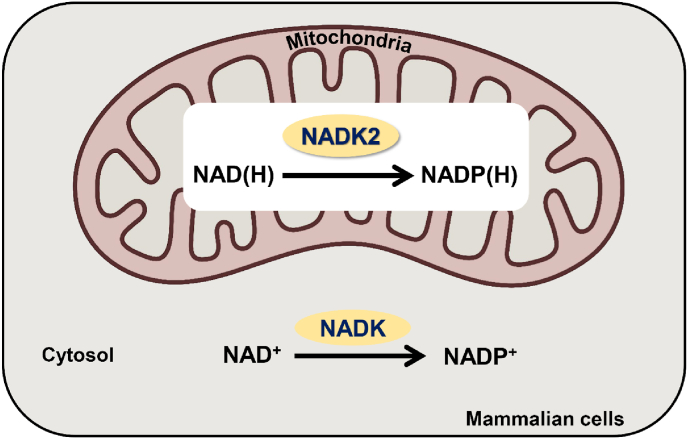
Nicotinamide adenine dinucleotide phosphate (NADP), a co-enzyme and an electron carrier, plays crucial roles in numerous biological functions, including cellular metabolism and antioxidation. Because NADP is subcellular-membrane impermeable, eukaryotes compartmentalize NAD kinases (NADKs), the NADP biosynthetic enzymes. Mitochondria are fundamental organelles for energy production through oxidative phosphorylation. Ten years after the discovery of the mitochondrial NADK (known as MNADK or NADK2), a significant amount of knowledge has been obtained regarding its functions, mechanism of action, human biology, mouse models, crystal structures, and post-translation modifications. NADK2 phosphorylates NAD(H) to generate mitochondrial NADP(H). NADK2-deficient patients suffered from hyperlysinemia, elevated plasma C10:2-carnitine (due to the inactivity of relevant NADP-dependent enzymes), and neuronal development defects. Nadk2-deficient mice recapitulate key features of NADK2-deficient patients, including metabolic and neuronal abnormalities. Crystal structures of human NADK2 show a dimer, with the NADP+-binding site located at the dimer interface. NADK2 activity is highly regulated by post-translational modifications, including S188 phosphorylation, K76 and K304 acetylation, and C193 S-nitrosylation; mutations in each site affect NADK2 activity and function. In mice, hepatic Nadk2 functions as a major metabolic regulator upon increased energy demands by regulating sirtuin 3 activity and fatty acid oxidation. Hopefully, future research on NADK2 will not only elucidate its functional roles in health and disease but will also pave the way for novel therapeutics for both rare and common diseases, including NADK2 deficiency and metabolic syndrome.
Keywords: Antioxidation; MNADK; Mitochondria; NAD; NADK; NADK2; NADP.
Copyright © 2023 The Authors. Published by Elsevier B.V. All rights reserved.
Conflict of interest statement
Declaration of competing interest The authors declare that they have no known competing financial interests or personal relationships that could have appeared to influence the work reported in this paper.
Figures


Similar articles
-
Structure of human NADK2 reveals atypical assembly and regulation of NAD kinases from animal mitochondria.Proc Natl Acad Sci U S A. 2022 Jun 28;119(26):e2200923119. doi: 10.1073/pnas.2200923119. Epub 2022 Jun 21. Proc Natl Acad Sci U S A. 2022. PMID: 35733246 Free PMC article.
-
The mitochondrial NAD kinase functions as a major metabolic regulator upon increased energy demand.Mol Metab. 2022 Oct;64:101562. doi: 10.1016/j.molmet.2022.101562. Epub 2022 Aug 6. Mol Metab. 2022. PMID: 35944895 Free PMC article.
-
Crystal structure of human NADK2 reveals a dimeric organization and active site occlusion by lysine acetylation.Mol Cell. 2022 Sep 1;82(17):3299-3311.e8. doi: 10.1016/j.molcel.2022.06.026. Epub 2022 Jul 21. Mol Cell. 2022. PMID: 35868311 Free PMC article.
-
MNADK, a Long-Awaited Human Mitochondrion-Localized NAD Kinase.J Cell Physiol. 2015 Aug;230(8):1697-701. doi: 10.1002/jcp.24926. J Cell Physiol. 2015. PMID: 25641397 Review.
-
Molecular properties and regulation of NAD+ kinase (NADK).Redox Biol. 2023 Feb;59:102561. doi: 10.1016/j.redox.2022.102561. Epub 2022 Dec 5. Redox Biol. 2023. PMID: 36512915 Free PMC article. Review.
Cited by
-
Pathobiochemistry of Aging and Neurodegeneration: Deregulation of NAD+ Metabolism in Brain Cells.Biomolecules. 2024 Dec 6;14(12):1556. doi: 10.3390/biom14121556. Biomolecules. 2024. PMID: 39766263 Free PMC article. Review.
-
Myeloma mesenchymal stem cells' bioenergetics afford a novel selective therapeutic target.Oncogenesis. 2025 Apr 11;14(1):9. doi: 10.1038/s41389-025-00554-5. Oncogenesis. 2025. PMID: 40216736 Free PMC article.
-
A 4D transcriptomic map for the evolution of multiple sclerosis-like lesions in the marmoset brain.bioRxiv [Preprint]. 2023 Sep 27:2023.09.25.559371. doi: 10.1101/2023.09.25.559371. bioRxiv. 2023. Update in: Science. 2025 Feb 28;387(6737):eadp6325. doi: 10.1126/science.adp6325. PMID: 37808784 Free PMC article. Updated. Preprint.
-
NADK tetramer defective mutants affect lung cancer response to chemotherapy via controlling NADK activity.Genes Dis. 2025 Jan 7;12(4):101510. doi: 10.1016/j.gendis.2024.101510. eCollection 2025 Jul. Genes Dis. 2025. PMID: 40330153 Free PMC article.
-
Mitochondrial Dysfunction in Heart Failure: From Pathophysiological Mechanisms to Therapeutic Opportunities.Int J Mol Sci. 2024 Feb 25;25(5):2667. doi: 10.3390/ijms25052667. Int J Mol Sci. 2024. PMID: 38473911 Free PMC article. Review.
References
-
- Ying W. NAD+/NADH and NADP+/NADPH in cellular functions and cell death: regulation and biological consequences. Antioxid Redox Signal. 2008;10:179–206. - PubMed
MeSH terms
Substances
Grants and funding
LinkOut - more resources
Full Text Sources
