Review
. 2023 Mar;1870(3):119432.
doi: 10.1016/j.bbamcr.2023.119432. Epub 2023 Jan 21.

Surveying the landscape of emerging and understudied cell death mechanisms

Affiliations
Review

Surveying the landscape of emerging and understudied cell death mechanisms

Logan Leak et al. Biochim Biophys Acta Mol Cell Res. 2023 Mar.

Abstract

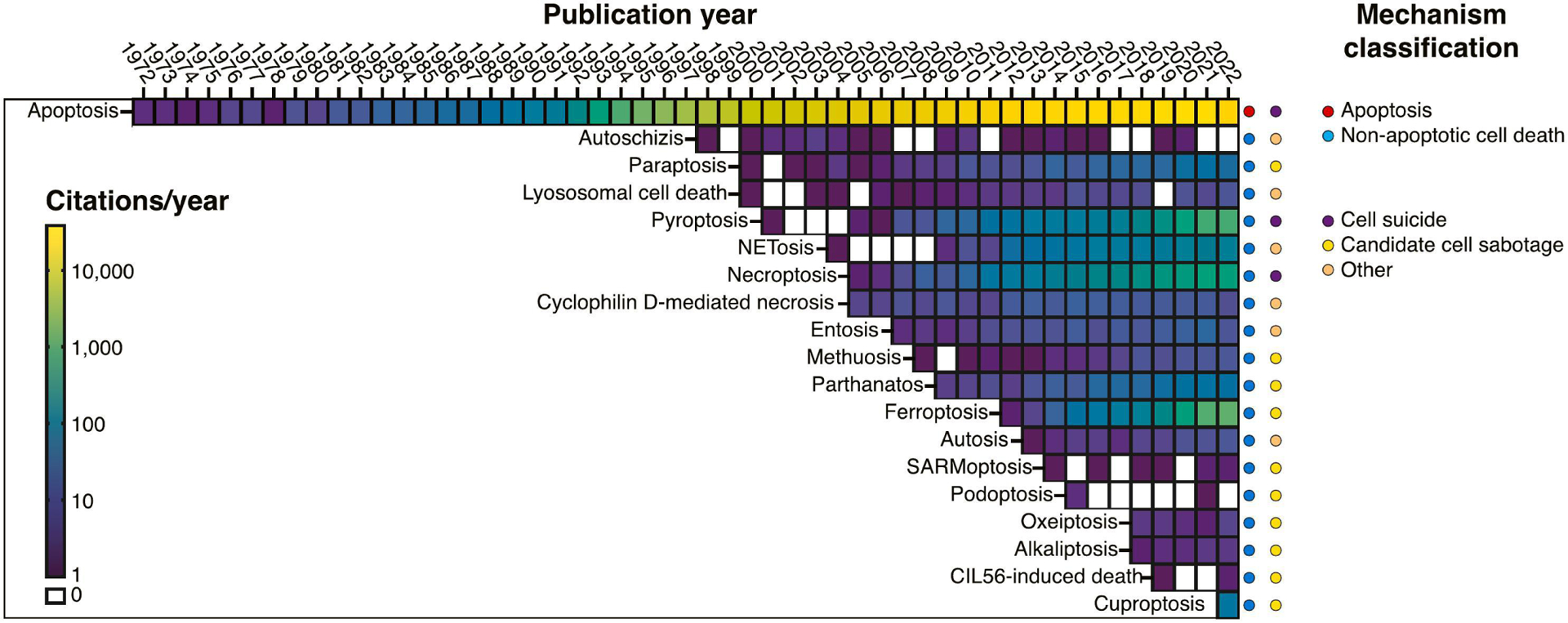
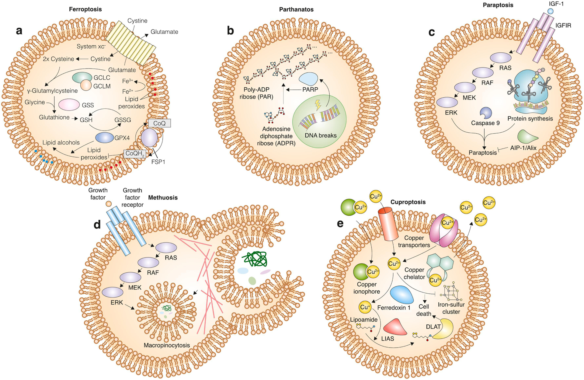
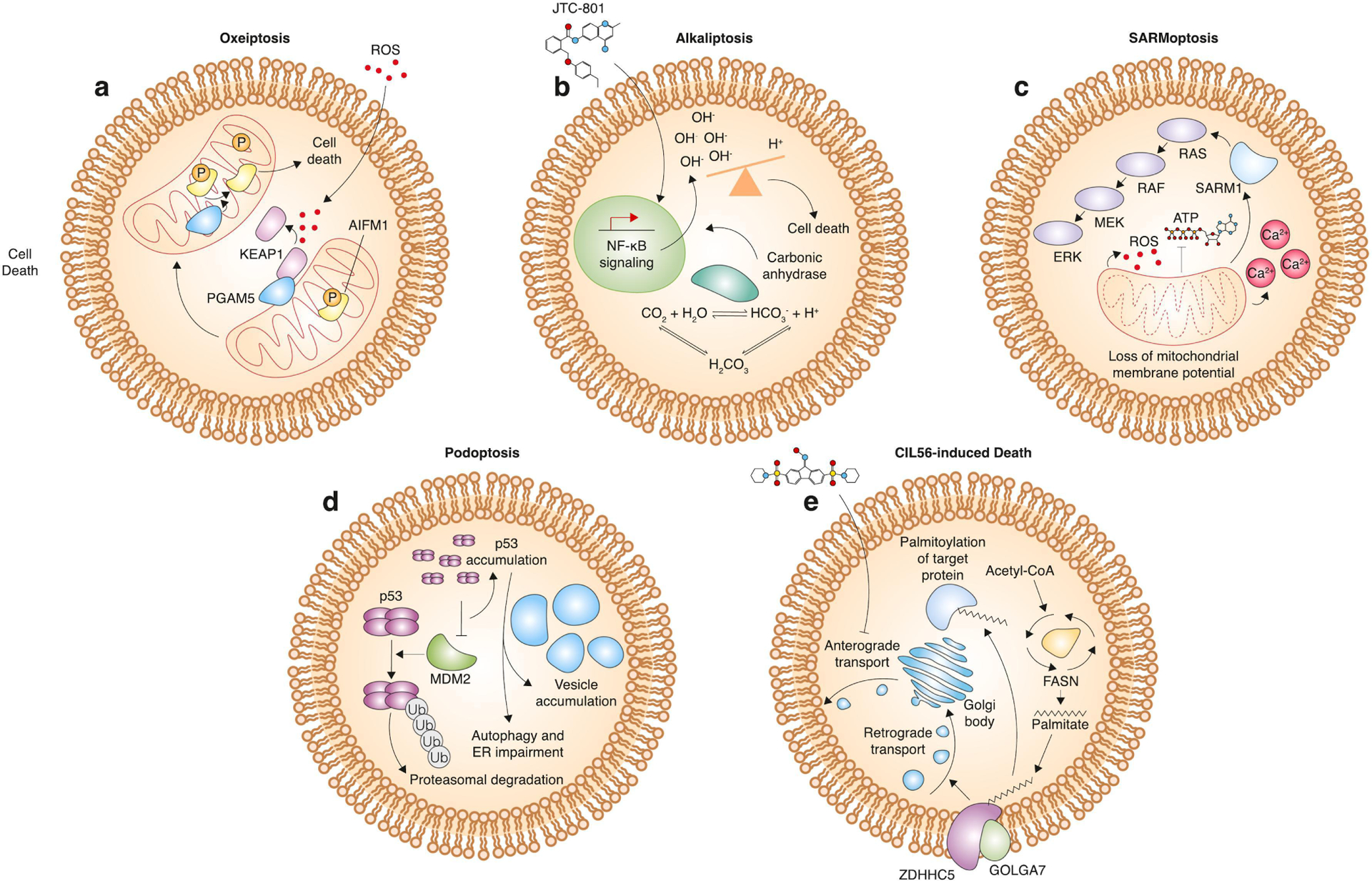
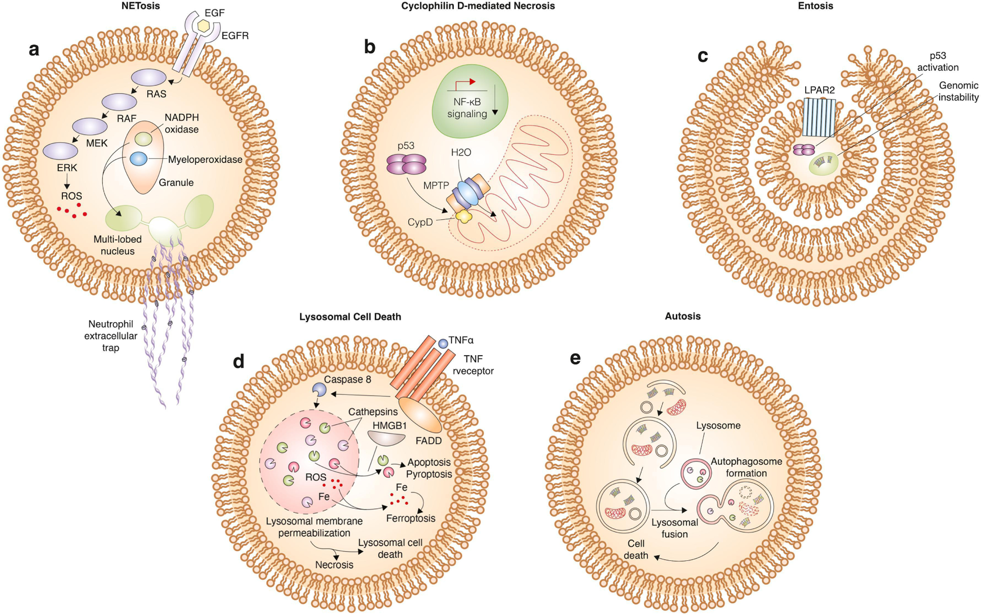
Cell death can be a highly regulated process. A large and growing number of mammalian cell death mechanisms have been described over the past few decades. Major pathways with established roles in normal or disease biology include apoptosis, necroptosis, pyroptosis and ferroptosis. However, additional non-apoptotic cell death mechanisms with unique morphological, genetic, and biochemical features have also been described. These mechanisms may play highly specialized physiological roles or only become activated in response to specific lethal stimuli or conditions. Understanding the nature of these emerging and understudied mechanisms may provide new insight into cell death biology and suggest new treatments for diseases such as cancer and neurodegeneration.

Keywords: Apoptosis; Ferroptosis; Necroptosis; Necrosis; Non-apoptotic cell death; Pyroptosis; ROS.


Conflict of interest statement

Declaration of competing interest The authors declare the following financial interests/personal relationships which may be considered as potential competing interests: Scott Dixon reports financial support was provided by Stanford University. Scott Dixon reports a relationship with Stanford University that includes: funding grants. S.J.D. is a co-founder of Prothegen Inc., a member of the scientific advisory board for Ferro Therapeutics and Hillstream BioPharma, and an inventor on patents related to ferroptosis.

Figures

Figure 1.
Figure 1.. Cell death mechanism citations over time.
Heatmap representing the number of citations reported in PubMed for different cell death mechanisms discussed in the review, arranged by year of first mention. Note that the first mention of a named pathway in PubMed in some cases comes years after the first observations of the mechanism, before it is recognized as unique and named. Each cell in the heatmap represents the number of PubMed citations mentioning each mechanism per calendar year. Years without any citations are white. On the right, cell death mechanisms are categorized as apoptotic or non-apoptotic, and sub-categorized as suicide, sabotage, or mixed/other mechanisms. This sub-classification is somewhat arbitrary and may shift over time as more is learned.
Figure 2.
Figure 2.. Cell suicide mechanisms.
Mechanism of apoptosis, necroptosis and pyroptosis. Apoptosis can be triggered by the extrinsic pathway involving binding of a ligand to its corresponding death receptor, caspase 8 activation, BH3 interacting domain death agonist (BID) cleavage into truncated BID (tBID), mitochondrial outer membrane permeabilization (MOMP), cytochrome C release, and apoptosome formation. Alternatively, the intrinsic pathway involves incorporation of signals that converge on the B cell lymphoma 2 (BCL2) family of proteins, which is comprised of pro- and anti-apoptotic proteins. When the pro-apoptotic proteins overcome inhibition by the anti-apoptotic proteins, MOMP occurs leading to formation of the apoptosome. Executioner caspase activation occurs following both extrinsic and intrinsic pathways and causes hallmarks of apoptosis including nuclear fragmentation, phosphatidyl serine exposure, and apoptotic body formation through blebbing occur. The mechanism of pyroptosis is distinct. Pathogen-associated molecular patterns (PAMPs) and damage-associated molecular patterns (DAMPs) bind to toll-like receptor 4 (TLR4) to activate specific caspases (1 and 4/5), which initiate the pyroptotic cascade. These caspases cleave gasdermin D into two fragments, with the N-terminal fragment translocating to the plasma membrane to create a pore through which processed interleukin 1 beta (IL-1β) can pass. The mechanism of necroptosis is likewise distinct from apoptosis and pyroptosis. Tumor necrosis factor alpha (TNFα) or other pro-death ligands bind to cell surface receptors to activate “complex I”, which includes TNF receptor type 1-associated death domain (TRADD), cellular inhibitor of apoptosis protein 1 (cIAP), and receptor interacting protein kinase 1 (RIPK1). Cylindromatosis lysine 63 deubiquitase (CYLD)/TNF alpha-induced protein 3 (TNFAIP3) catalyzes the deubiquitylation of RIPK1, which enables the formation of “complex II”. Complex II includes RIPK1, TRADD, Fas-associated death domain (FADD), and caspase 8. When caspase 8 is inactive, RIPK3 associates with RIPK1 to promote phosphorylation and subsequent polymerization of mixed lineage kinase domain like pseudokinase (MLKL). Polymerized MLKL localizes to the plasma membrane where it forms a pore through which DAMPs and cytokines are released.
Figure 3.
Figure 3.. Mechanisms of cell death that fit into the cell suicide/sabotage dichotomy.
a, mechanism of ferroptosis. The cystine/glutamate antiporter system xc normally allows for entry of cystine into the cell, where it can be reduced into cysteine and incorporated into the antioxidant tripeptide glutathione. Glutathione is a cofactor for the phospholipid hydroperoxidase glutathione peroxidase 4 (GPX4), which catalyzes the conversion of potentially toxic lipid peroxides into non-toxic lipid alcohols. In parallel, the oxidoreductase ferroptosis suppressor protein 1 (FSP1) can catalyze the synthesis of reduced electron carriers such as coenzyme Q10 (CoQ10-H2), which in turn inhibit lipid reactive oxygen species (ROS) spread at the plasma membrane. Inhibition of these lipid peroxide defenses leads to ferroptosis. b, mechanism of parthanatos. DNA breaks lead to activation of poly (adenosine diphosphate-ribose) polymerase (PARP), which then catalyzes the polymerization of adenosine diphosphate (ADP) ribose into poly (adenosine diphosphate-ribose) (PAR), which leads to death induction. c, mechanism of paraptosis. Activation of insulin-like growth factor receptor 1 (IGF1R) by insulin-like growth factor (IGF-1) leads to downstream activation of the mitogen-activated protein kinase (MAPK) pathway. The MAPK pathway activates paraptosis in a protein synthesis- and caspase 9-dependent manner. This death pathway is opposed by actin-integrating protein 1 (AIP-1)/apoptosis-linked gene 2-interacting protein X (Alix). d, mechanism of methuosis. Growth factors bind to their respective receptors to activate the MAPK pathway. Increased signaling through this pathway induces micropinocytosis of extracellular material. Excessive micropinocytosis leads to dramatic vacuolization that physically detaches the cell from its substrate and leads to death induction. e, mechanism of cuproptosis. Intracellular copper levels are modulated by a variety of factors. Copper transporters shuttle copper in either direction across the membrane. Copper ionophores bind to and bring copper ions into the cell across the membrane, and copper chelators sequester copper and decrease cytosolic levels. Intracellular copper is oxidized by ferredoxin 1 and then serves as a co-factor for lipoic acid synthase (LIAS)-mediated lipoylation of target proteins. Lipoylation of dihydrolipoamide S-acetyltransferase (DLAT) and copper-dependent inhibition of iron-sulfur cluster formation leads to cuproptosis.
Figure 4.
Figure 4.. Mechanisms of cell death that fit into the cell suicide/sabotage dichotomy (continued).
a, mechanism of oxeiptosis. Excessive ROS leads to dissociation of kelch like ECH associated protein 1 (KEAP1) from phosphoglycerate mutase 5 (PGAM5) and subsequent release of PGAM5 from the mitochondria. PGAM5 can then act as a phosphatase to remove phosphates from apoptosis-inducing factor (AIFM1), which then leads to death induction. b, mechanism of alkaliptosis. JTC-801 leads to increased NF-κB pathway activity, which decreases expression of carbonic anhydrase. This leads to an increase in intracellular pH and cell death. c, mechanism of SARMoptosis. Compromising of mitochondrial membrane potential leads to ROS accumulation, depletion of intracellular ATP, accumulation of calcium, and sterile alpha and Toll/interleukin receptor (TIR) motif containing 1 (SARM) activation. SARM can activate the MAPK pathway, which in turn results in cell death. d, mechanism of podoptosis. Inhibition of the E3 ubiquitin ligase mouse double minute 2 (MDM2) leads to accumulation of the p53 tetramer. p53 accumulation causes vesicle accumulation and impairment of autophagy and normal endoplasmic reticulum homeostasis, leading to cell death. e, Caspase-independent lethal (CIL56)-induced cell death. CIL56 inhibits anterograde transport from the Golgi body to the plasma membrane. Zinc finger DHHC-type palmitoyltransferase 5 (ZDHHC5) and golgin A7 (GOLGA7) form a protein S-acylation complex that promotes retrograde transport from the plasma membrane to the Golgi body. This process is dependent upon palmitate, a 16-carbon saturated fatty acid.
Figure 5.
Figure 5.. Mechanisms of cell death that do not fit into the cell suicide/sabotage dichotomy.
a, mechanism of NETosis. Activation of the MAPK pathway by binding of epidermal growth factor (EGF) to its associated receptor leads to ROS accumulation. ROS stimulates the release of nicotinamide adenine dinucleotide phosphate (NADPH) oxidase and myeloperoxidase from granules within the neutrophil. Translocation of these proteins to the multi-lobed nucleus results in release of nuclear contents, including DNA and histones, into the extracellular space where they can interact with target cells to induce cell death. b, mechanism of Cyclophilin D-mediated necrosis. The mitochondria permeability transition pore (MPTP), upon activation involving binding of CypD, results in permeabilization of the outer mitochondrial membrane. This releases water into the mitochondria that causes it to burst open and induce cell death. This form of cell death is associated with decreased nuclear factor kappa-light-chain-enhancer of activated B cells (NF-κB signaling). c, mechanism of entosis. Cell cannibalism or the cell-in-cell phenotype is activated by close contact of cells and formation of adherens junctions. In entosis, one cell is completely engulfed within another, and the process is activated by G-protein coupled lysophosphatidic acid receptor 2 (LPAR2) activation, p53 activation, and genomic instability. The cell can be recycled back to the plasma membrane or killed by enzymes within the lysosome upon fusion of the cell-containing vesicle with the lysosome. d, mechanism of lysosomal cell death. TNFα binds to its receptor to activate FADD and caspase 8. Caspase 8 induces lysosomal membrane permeabilization and subsequent release of cathepsins, ROS, and iron into the cytosol. High mobility group box 1 (HMGB1) facilitates the release of cathepsins specifically, which leads to induction of apoptosis or pyroptosis. Alternatively, cytosolic ROS accumulation can lead to the induction of ferroptosis. In addition, cell death by necrosis or an independent lysosomal cell death pathway can ensue. e, mechanism of autosis. Activation of autophagy leads to recycling of cellular components including proteins, nucleic acids, and organelles. Fusion with the lysosome leads to degradation of these components, and excessive activation of this pathway leads to cell death.

Similar articles

Cited by

References

    1. Mazzarello P, A unifying concept: the history of cell theory, Nat Cell Biol, 1 (1999) E13–E15. - PubMed
    1. Vogt C, Untersuchungen über die Entwicklungsgeschichte der Geburtshelferkrœte (Alytes obstetricans), Jent & Gassmann, 1842.
    1. Majno G, Joris I, Apoptosis, oncosis, and necrosis An overview of cell death, Am J Pathol, 146 (1995) 3–15. - PMC - PubMed
    1. Kerr JFR, Wyllie AH, Currie AR, Apoptosis: A Basic Biological Phenomenon with Wideranging Implications in Tissue Kinetics, Br J Cancer, 26 (1972) 239–257. - PMC - PubMed
    1. Tata JR, Requirement for RNA and protein synthesis for induced regression of the tadpole tail in organ culture, Developmental Biology, 13 (1966) 77–94. - PubMed

Publication types

LinkOut - more resources