. 2023 Mar 28;100(13):e1418-e1432.
doi: 10.1212/WNL.0000000000206820. Epub 2023 Jan 23.

Tumefactive Demyelination in MOG Ab-Associated Disease, Multiple Sclerosis, and AQP-4-IgG-Positive Neuromyelitis Optica Spectrum Disorder

Affiliations

Tumefactive Demyelination in MOG Ab-Associated Disease, Multiple Sclerosis, and AQP-4-IgG-Positive Neuromyelitis Optica Spectrum Disorder

Laura Cacciaguerra et al. Neurology. .

Abstract

Background and objectives: Studies on tumefactive brain lesions in myelin oligodendrocyte glycoprotein-immunoglobulin G (IgG)-associated disease (MOGAD) are lacking. We sought to characterize the frequency clinical, laboratory, and MRI features of these lesions in MOGAD and compare them with those in multiple sclerosis (MS) and aquaporin-4-IgG-positive neuromyelitis optica spectrum disorder (AQP4+NMOSD).

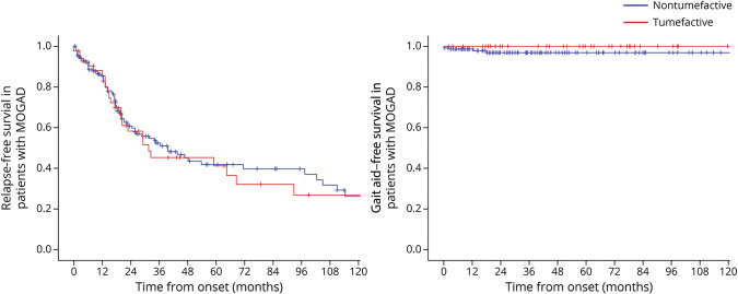
Methods: We retrospectively searched 194 patients with MOGAD and 359 patients with AQP4+NMOSD with clinical/MRI details available from the Mayo Clinic databases and included those with ≥1 tumefactive brain lesion (maximum transverse diameter ≥2 cm) on MRI. Patients with tumefactive MS were identified using the Mayo Clinic medical record linkage system. Binary multivariable stepwise logistic regression identified independent predictors of MOGAD diagnosis; Cox proportional regression models were used to assess the risk of relapsing disease and gait aid in patients with tumefactive MOGAD vs those with nontumefactive MOGAD.

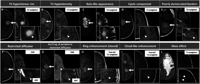
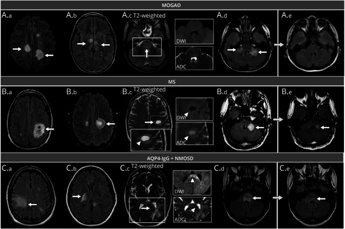
Results: We included 108 patients with tumefactive demyelination (MOGAD = 43; AQP4+NMOSD = 16; and MS = 49). Tumefactive lesions were more frequent among those with MOGAD (43/194 [22%]) than among those with AQP4+NMOSD (16/359 [5%], p < 0.001). Risk of relapse and need for gait aid were similar in tumefactive and nontumefactive MOGAD. Clinical features more frequent in MOGAD than in MS included headache (18/43 [42%] vs 10/49 [20%]; p = 0.03) and somnolence (12/43 [28%] vs 2/49 [4%]; p = 0.003), the latter also more frequent than in AQP4+NMOSD (0/16 [0%]; p = 0.02). The presence of peripheral T2-hypointense rim, T1-hypointensity, diffusion restriction (particularly an arc pattern), ring enhancement, and Baló-like or cystic appearance favored MS over MOGAD (p ≤ 0.001). MRI features were broadly similar in MOGAD and AQP4+NMOSD, except for more frequent diffusion restriction in AQP4+NMOSD (10/15 [67%]) than in MOGAD (11/42 [26%], p = 0.005). CSF analysis revealed less frequent positive oligoclonal bands in MOGAD (2/37 [5%]) than in MS (30/43 [70%], p < 0.001) and higher median white cell count in MOGAD than in MS (33 vs 6 cells/μL, p < 0.001). At baseline, independent predictors of MOGAD diagnosis were the presence of somnolence/headache, absence of T2-hypointense rim, lack of T1-hypointensity, and no diffusion restriction (Nagelkerke R 2 = 0.67). Tumefactive lesion resolution was more common in MOGAD than in MS or AQP4+NMOSD and improved model performance.

Discussion: Tumefactive lesions are frequent in MOGAD but not associated with a worse prognosis. The clinical, MRI, and CSF attributes of tumefactive MOGAD differ from those of tumefactive MS and are more similar to those of tumefactive AQP4+NMOSD with the exception of lesion resolution, which favors MOGAD.


Conflict of interest statement

L. Cacciaguerra received speaker and consultant honoraria from ACCMED, Roche, BMS Celgene, and Sanofi. P. Morris reports no disclosures. W.O. Tobin has received research funding from Mallinckrodt Inc., the Mayo Clinic Center for Multiple Sclerosis and Autoimmune Neurology, and the NIH (grant R01NS113803 and R01NS121928) outside the submitted work; he has received honoraria from Neurology Live and has coedited Mayo Clinic Cases in Neuroimmunology. J.J. Chen served as consultant for Roche and UCB. S.A. Banks, P. Elsbernd, and V. Redenbaugh report no disclosures. J.-M. Tillema is associate editor for the Journal of Child Neurology. F. Montini and E. Sechi report no disclosures. A.S. Lopez-Chiriboga has served on advisory boards for Genentech and Horizon Therapeutics. N.L. Zalewski and Y. Guo report no disclosures. M. Assunta Rocca received speaker honoraria from Bayer, Biogen, Bristol Myers Squibb, Celgene, Genzyme, Merck Serono, Novartis, Roche, and Teva and receives research support from the MS Society of Canada and Fondazione Italiana Sclerosi Multipla. M. Filippi is editor-in-chief of the Journal of Neurology, associate editor of Human Brain Mapping, associate editor of Radiology, and associate editor of Neurological Sciences; received compensation for consulting services and/or speaking activities from Almiral, Alexion, Bayer, Biogen, Celgene, Eli Lilly, Genzyme, Merck-Serono, Novartis, Roche, Sanofi, Takeda, and Teva Pharmaceutical Industries; and receives research support from Biogen Idec, Merck-Serono, Novartis, Roche, Teva Pharmaceutical Industries, Italian Ministry of Health, Fondazione Italiana Sclerosi Multipla, and ARiSLA (Fondazione Italiana di Ricerca per la SLA). S.J. Pittock reports grants, personal fees, and nonfinancial support from Alexion Pharmaceuticals, Inc.; grants, personal fees, nonfinancial support, and other support from MedImmune, Inc./Viela Bio, Inc.; and personal fees for consulting from Genentech/Roche; he has a patent, patent # 8,889,102 (application #12-678350, Neuromyelitis Optica Autoantibodies as a Marker for Neoplasia)—issued and a patent, patent # 9,891,219B2 (application #12-573942, Methods for Treating Neuromyelitis Optica [NMO] by Administration of Eculizumab to an individual that is Aquaporin-4 [AQP4]–IgG Autoantibody positive)—issued. C.F. Lucchinetti received grants from the National Institute of Health, National Multiple Sclerosis Society, National Institute of Neurological Disorders and Stroke, Kingsland Foundation, Biogen Idec. E.P. Flanagan has served on advisory boards for Alexion, Genentech, and Horizon Therapeutics, has received speaker honoraria from Pharmacy Times, and received royalties from UpToDate. Dr. Flanagan was a site primary investigator in a randomized clinical trial on Inebilizumab in neuromyelitis optica spectrum disorder run by Medimmune/Viela-Bio/Horizon Therapeutics, has received funding from the NIH (R01NS113828), and is a member of the medical advisory board of the MOG project. Dr. Flanagan is an editorial board member of the Journal of the Neurological Sciences and Neuroimmunology Reports, and a patent has been submitted on DACH1-IgG as a biomarker of paraneoplastic autoimmunity. Go to Neurology.org/N for full disclosures.

Figures

Figure 1
Figure 1. Pictorial Overview of the MRI Features Evaluated in This Study, With Corresponding Definitions
Lesions are shown by arrows and are displayed on axial T2-FLAIR (left half of each image) and on the most appropriate sequence for the evaluation of each specific feature, indicated by an arrowhead (right half of each image). Exceptions are images E and J, where the second sequence (T2-weighted image in E, and T1-weighted image in J) is reported for descriptive purposes only because the T2-FLAIR sequence is suitable for the visualization of those features. For image I, T2-FLAIR image was unavailable and therefore substituted with T2-weighted sequence. Features were defined as follows: (A) T2-hypointense rim: rim-shaped relative T2-hypointensity compared with the T2-hyperintensity of the lesion center and surrounding edema; (B) T1-hypointensity: hypointensity on T1-weighted images compared with the normal-appearing white matter; (C) Baló-like appearance: the presence of ≥2 concentric rings or a pattern of alternating bands of signal intensity on any MRI sequence; (D) cystic component: T2-weighted or T1-weighted signal of similar intensity to CSF; (E) poorly demarcated borders: fluffy appearance on T2-FLAIR or T2-weighted sequences; (F) restricted diffusion: DWI hyperintensity with corresponding hypointensity on the ADC map; (G) arc/ring of peripheral restricted diffusion: dark peripheral ADC arc or ring with a brighter center; (H) ring enhancement (closed ring shown in this figure): circular border of enhancement, closed if complete, otherwise open; (I) cloud-like enhancement: multiple patchy enhancement with ill-defined margins; (J) mass effect: lesion exerting a secondary effect on adjacent structures such as sulci, ventricles, or midline. ADC = apparent diffusion coefficient map; DWI = diffusion-weighted images; FLAIR = fluid-attenuated inversion recovery; Gd = gadolinium.
Figure 2
Figure 2. Long-term Outcomes of Patients With MOGAD With Tumefactive Lesions
Kaplan-Meier curves of survival probability estimates of not developing relapse or reaching the EDSS disability milestone of 6 in patients with MOGAD up to 120 months from disease onset, according to the presence of tumefactive lesions. EDSS = Expanded Disability Status Scale; IgG = immunoglobulin G; MOGAD = myelin oligodendrocyte glycoprotein-IgG–associated disease.
Figure 3
Figure 3. Examples of Tumefactive Brain Lesions and Relevant MRI Features for the Differential Diagnosis of Patients With MOGAD, MS, and AQP4+NMOSD
Unless otherwise specified, lesions are shown on axial T2-fluid-attenuated inversion recovery (T2-FLAIR). Tumefactive lesions are indicated by white arrows, while specific features are indicated by arrowheads. Panel A (MOGAD): tumefactive lesions in the bilateral hemispheric white matter (A.a, arrows); bilateral tumefactive lesions in the corticospinal tract and thalami (A.b, arrows); tumefactive lesion involving the entire pons (A.c, arrow) without evidence of T2-hypointense rim, diffusion restriction (i.e., no DWI hyperintensity), and no ADC map hypointensity (A.c); bilateral tumefactive lesions in the middle cerebellar peduncles (A.d, arrows), with complete resolution at follow-up MRI (A.e). Panel B (MS): Baló-like tumefactive lesions in the hemispheric white matter (B.a, arrow) and periventricular white matter (B.b, arrow); tumefactive lesion located in the hemispheric white matter, showing a complete T2-hypointense rim (arrowhead) with a corresponding ring of peripheral diffusion restriction, as indicated by the hyperintensity in DWI (arrowhead) and hypointensity on ADC map (arrowhead) (B.c); tumefactive lesion in the left middle cerebellar peduncle (B.d, arrow) undergoing volumetric reduction, but still present at follow-up MRI (B.e, arrow). Panel C (AQP4+NMOSD): tumefactive lesions in the hemispheric white matter (C.a, arrow), corticospinal tract (C.b, arrow); tumefactive periventricular lesion extensively involving the splenium of the corpus callosum in an “arch-bridge” pattern (C.c, arrow); the lesion does not show any T2-hypointense rim and is characterized by DWI hyperintensity of the splenium (arrowhead) with focal diffusion restriction (i.e., corresponding hypointensity on the ADC map, arrowhead); tumefactive lesion involving the entire pons (C.d, arrow), showing residual mild hyperintensity near the fourth ventricle (C.e, arrow). ADC = apparent diffusion coefficient map; AQP4+NMOSD = aquaporin-4-IgG–positive neuromyelitis optica spectrum disorders; DWI = diffusion-weighted images; IgG = immunoglobulin G; MOGAD = myelin oligodendrocyte glycoprotein-IgG–associated disease; MS = multiple sclerosis.

References

    1. Reindl M, Waters P. Myelin oligodendrocyte glycoprotein antibodies in neurological disease. Nat Rev Neurol. 2019;15(2):89-102. doi:10.1038/s41582-018-0112-x. - DOI - PubMed
    1. Jarius S, Paul F, Aktas O, et al. . MOG encephalomyelitis: international recommendations on diagnosis and antibody testing. J Neuroinflammation. 2018;15(1):134. doi:10.1186/s12974-018-1144-2. - DOI - PMC - PubMed
    1. Hardy TA, Chataway J. Tumefactive demyelination: an approach to diagnosis and management. J Neurol Neurosurg Psychiatry. 2013;84(9):1047-1053. doi:10.1136/jnnp-2012-304498. - DOI - PubMed
    1. Lucchinetti CF, Gavrilova RH, Metz I, et al. . Clinical and radiographic spectrum of pathologically confirmed tumefactive multiple sclerosis. Brain. 2008;131(7):1759-1775. doi:10.1093/brain/awn098. - DOI - PMC - PubMed
    1. Shu Y, Long Y, Wang S, et al. . Brain histopathological study and prognosis in MOG antibody-associated demyelinating pseudotumor. Ann Clin Transl Neurol. 2019;6(2):392-396. doi:10.1002/acn3.712. - DOI - PMC - PubMed

Publication types