. 2023 Jan 23;13(1):1256.
doi: 10.1038/s41598-022-27018-9.

Colitis in a transgenic mouse model of autoimmune uveitis may be induced by neoantigen presentation in the bowel

Affiliations

Colitis in a transgenic mouse model of autoimmune uveitis may be induced by neoantigen presentation in the bowel

C Mölzer et al. Sci Rep. .

Abstract

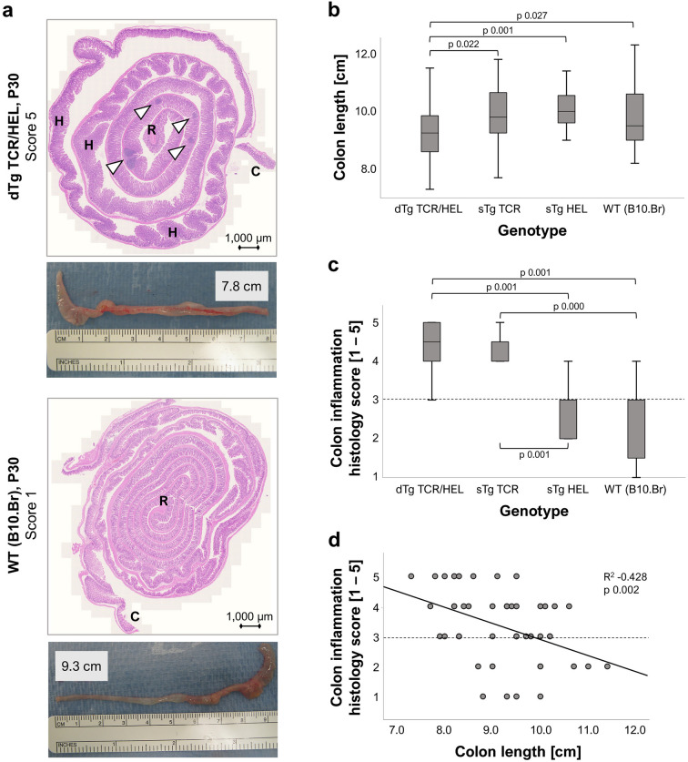
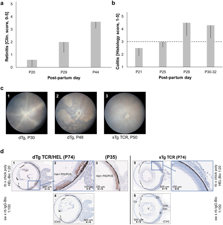
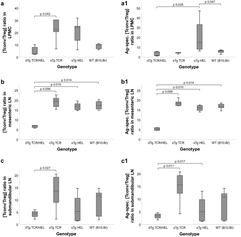
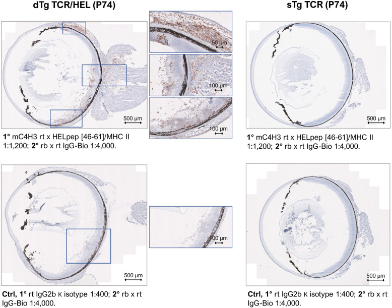
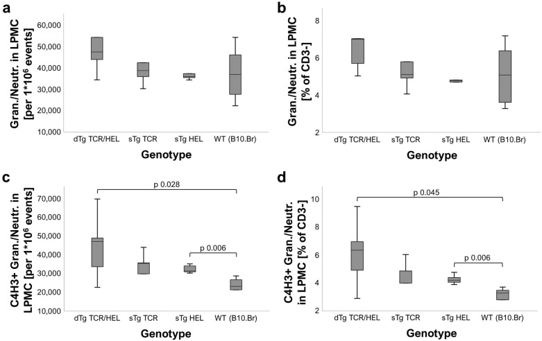
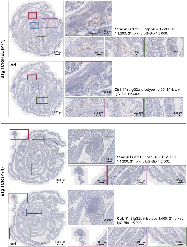
Undifferentiated uveitis (intraocular inflammation, IOI) is an idiopathic sight-threatening, presumed autoimmune disease, accountable for ~ 10% of all blindness in the developed world. We have investigated the association of uveitis with inflammatory bowel disease (IBD) using a mouse model of spontaneous experimental autoimmune uveoretinitis (EAU). Mice expressing the transgene (Tg) hen egg lysozyme (HEL) in the retina crossed with 3A9 mice expressing a transgenic HEL-specific TCR spontaneously develop uveoretinitis at post-partum day (P)20/21. Double transgenic (dTg TCR/HEL) mice also spontaneously develop clinical signs of colitis at ~ P30 with diarrhoea, bowel shortening, oedema and lamina propria (LP) inflammatory cell infiltration. Single (s)Tg TCR (3A9) mice also show increased histological LP cell infiltration but no bowel shortening and diarrhoea. dTg TCR/HEL mice are profoundly lymphopenic at weaning. In addition, dTg TCR/HEL mice contain myeloid cells which express MHC Class II-HEL peptide complexes (MHCII-HEL), not only in the inflamed retina but also in the colon and have the potential for antigen presentation. In this model the lymphopenia and reduction in the absolute Treg numbers in dTg TCR/HEL mice is sufficient to initiate eye disease. We suggest that cell-associated antigen released from the inflamed eye can activate colonic HEL-specific T cells which, in a microbial micro-environment, not only cause colitis but feedback to amplify IOI.


Conflict of interest statement

The authors declare that the research was conducted in the absence of any commercial or financial relationships that could be construed as a potential conflict of interest. RC is a founder of MIROBio Ltd.

Figures

Figure 1
Figure 1
Mouse large intestines exhibit genotype-specific histological features. (a) H&E-stained colon section (Swiss roll) of dTg mice shows clear signs of inflammation (granulomatous colitis). Cardinal features are granulomas/crypt abscesses (white arrowheads) and crypt hyperplasia (H), along with bowel shortening and oedema. WT mice have longer bowels with normal appearance of mucosal epithelium. Abbreviations: R, rectum; C, caecum. (b) Colons of dTg mice are significantly shorter than those of all other genotypes; (c) Both dTg and sTg TCR colons have significantly more inflamed phenotypes as compared to the sTg HEL and WT mice. (d) Colon length and histological inflammation score are significantly correlated. Dashed lines indicate clinically irrelevant “background” colon inflammation. Non-parametric statistical procedures were used; significance scored at p ≤ 0.05. For each genotype group n = 10–13 colons were independently examined. Mice were aged P30 ± 3 days.
Figure 2
Figure 2
Colitis and retinitis (spontaneous EAU) have distinct clinical onsets. (a) Mice with EAU (n = 7–8, assessed independently) spontaneously develop uveitis from P20 with gradual worsening of inflammation until P44 when the disease wears off leaving behind irreversible retinal atrophy. (b) Clinical signs of colitis (i.e., diarrhoea, rectal prolapse) were observed in dTg mice (n = 4 per age-group, independently assessed) from around P30. Histological staining and subsequent scoring revealed the presence of colon inflammation after P25 (clinical scores between 1 and 3 were identified as the baseline range, dashed line). Error bars indicate sd. (c) Representative fundoscopy images of P30 dTg (1, vasculitis), P48 dTg (2, retinal atrophy), and P50 TCR eyes (3, normal retina) (n = 34 eye pairs/genotype group; independently assessed. (d) Eye sections of dTg aged mice (P74; n = 3, independently stained, three replicates) show transgenic expression of HEL protein in their photoreceptor layer (1, 2; brown staining). This is specific to this spontaneous EAU mouse model in which HEL is the uveitogenic antigen. Photoreceptor structure is better preserved in younger eyes (3). Note the absence of staining in control section (ctrl; 4) as well as sTg TCR eyes that are devoid of the retinal transgene (5, 6). Abbreviations: PIS/POS, photoreceptor layer inner and outer segment; Ch, choroid; R, retina; ON/OD, optic nerve/disc; V, vitreous; Co, cornea.
Figure 3
Figure 3
Mice with the dTg genotype have relatively lower (antigen-specific) [Tconv/Treg] cell ratios across different tissues. (a, a1) in LPMC (lamina propria mononucleated cells); (b, b1) in mesenteric lymph nodes (LN); and (c, c1) in submandibular (eye-draining) LN. dTg mice were found to have significantly lower [Tconv/Treg] quotients across all tissues as compared to their non-lymphopenic counterparts. The same was true with reference to antigen-specificity. Treg cells had been defined as CD4+ CD25+ FoxP3+, Tconv as CD4+ CD25± FoxP3-. Antigen-specific populations had been classified as Vbeta8.1/8.2+ . Ratios were calculated based on absolute cell numbers [n] acquired using flow cytometry. Non-parametric statistical procedures were used, significance taken as p ≤ 0.05. Per genotype group n = 5–7 mice (aged P30 ± 3) were used across 2–3 independent experiments.
Figure 4
Figure 4
MHCII-HEL complexes are exclusively present in retinas of dTg mice. The monoclonal C4H3 rat anti-chicken antibody towards HEL peptide [46–61] was used to stain MHCII-HEL complexes in eye sections. OCT sections were counterstained with Haematoxylin QS (Vector Laboratories, 2BScientific, UK). Left panel: P74 section from dTg mouse; note positive brown staining particularly in and around inflamed retinal tissue and uveal tract vs. control (faint background only). Right panel: staining was performed using sTg TCR eye sections. No staining was found in either sTg TCR test conditions. Per genotype group n = 3 sections were independently stained across three replicates.
Figure 5
Figure 5
MHCII-HEL staining positivity is significantly stronger in lamina propria mononucleated cells isolated from dTg colon. MHCII-HEL complexes were detected using flow cytometry in granulocytes/neutrophils in LPMC (lamina propria mononucleated cells) isolated from mouse colon (aged P30 ± 3). While no difference was found in either (a) total granulocyte/neutrophil numbers recorded, and (b) cell counts relative to all CD3- events, there were significantly more MHCII-HEL presenting cells [C4H3+; (c) and (d)] found in LPMC isolated from dTg mice as compared to all other genotypes (= background level staining). This result is indicative of potential for HEL antigen presentation capacity in colons of dTg mice. Cells had been defined as CD3- CD11b+ Gr1/Ly6+. Non-parametric statistical procedures were used, significance taken as p ≤ 0.05. Per genotype group n = 5–7 mice were used across 2–3 independent experiments.
Figure 6
Figure 6
MHCII-HEL complexes are exclusively present in colon of dTg mice. The monoclonal C4H3 rat anti-chicken antibody towards HEL peptide [46–61] was used to histologically stain MHCII-HEL complexes in colon Swiss roll sections. Sections were counterstained with Haematoxylin QS (Vector Laboratories, 2BScientific, UK). Top panel: P74 section from dTg mouse; note positive brown staining particularly in and around granulomas and erupting Peyer’s patches vs. control. Bottom panel: staining was performed using sTg TCR colon sections. Only faint background staining was found in either sTg TCR test conditions. Note: Unavoidable faint background staining results from the intrinsic abundance of inflammatory cells in the lamina propria. Per genotype group n = 3 sections were independently stained across three replicates.

Similar articles

Cited by

References

    1. Jabs DA. Epidemiology of uveitis. Ophthalmic Epidemiol. 2008;15:283–284. doi: 10.1080/09286580802478724. - DOI - PubMed
    1. Jabs DA, Nussenblatt RB, Rosenbaum JT. Standardization of uveitis nomenclature working, G. Standardization of uveitis nomenclature for reporting clinical data. Results of the First International Workshop. Am. J. Ophthalmol. 2005;140:509–516. doi: 10.1016/j.ajo.2005.03.057. - DOI - PMC - PubMed
    1. Nussenblatt RB. The natural history of uveitis. Int. Ophthalmol. 1990;14:303–308. doi: 10.1007/BF00163549. - DOI - PubMed
    1. Scherer, H. U., van der Woude, D. & Toes, R. E. From risk to chronicity: Evolution of autoreactive B cell and antibody responses in rheumatoid arthritis. Nat. Rev. Rheumatol. 1–13 (2022). - PubMed
    1. Forrester JV, Kuffova L, Dick AD. Autoimmunity, Autoinflammation and Infection in Uveitis. Am. J. Ophthalmol. 2018 doi: 10.1016/j.ajo.2018.02.019. - DOI - PubMed

Publication types

Substances