. 2023 Jan 23;6(1):86.
doi: 10.1038/s42003-023-04484-z.

TRIM24 controls induction of latent HIV-1 by stimulating transcriptional elongation

Affiliations

TRIM24 controls induction of latent HIV-1 by stimulating transcriptional elongation

Riley M Horvath et al. Commun Biol. .

Abstract

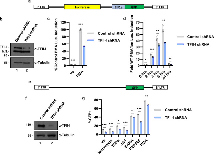
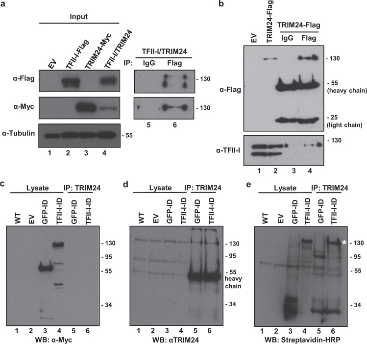
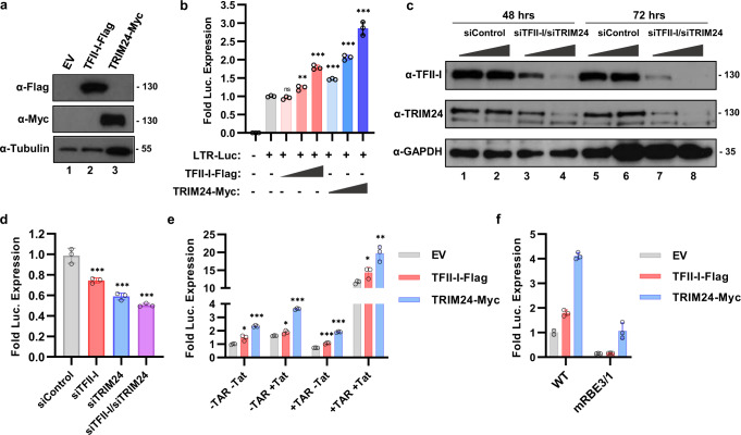
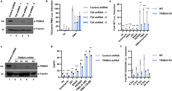
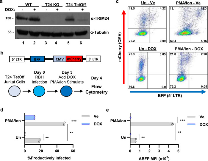
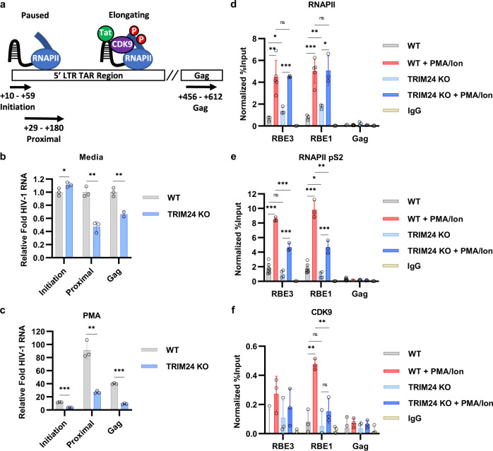
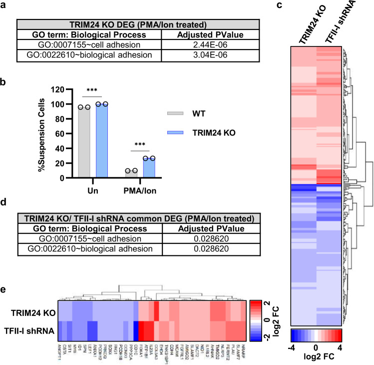
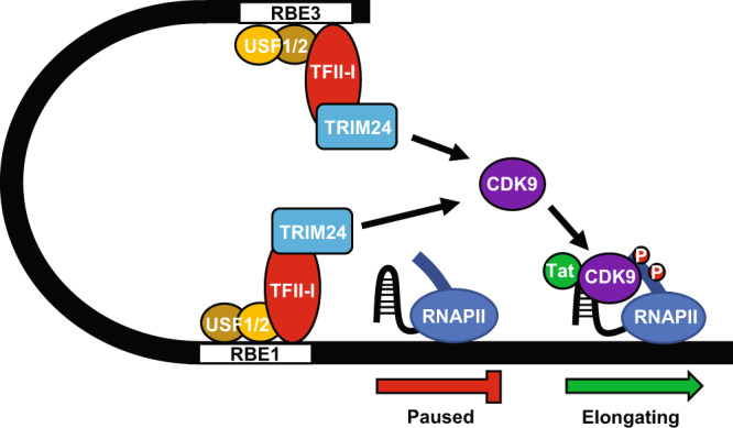
Binding of USF1/2 and TFII-I (RBF-2) at conserved sites flanking the HIV-1 LTR enhancer is essential for reactivation from latency in T cells, with TFII-I knockdown rendering the provirus insensitive to T cell signaling. We identified an interaction of TFII-I with the tripartite motif protein TRIM24, and these factors were found to be constitutively associated with the HIV-1 LTR. Similar to the effect of TFII-I depletion, loss of TRIM24 impaired reactivation of HIV-1 in response to T cell signaling. TRIM24 deficiency did not affect recruitment of RNA Pol II to the LTR promoter, but inhibited transcriptional elongation, an effect that was associated with decreased RNA Pol II CTD S2 phosphorylation and impaired recruitment of CDK9. A considerable number of genomic loci are co-occupied by TRIM24/TFII-I, and we found that TRIM24 deletion caused altered T cell immune response, an effect that is facilitated by TFII-I. These results demonstrate a role of TRIM24 for regulation of transcriptional elongation from the HIV-1 promoter, through its interaction with TFII-I, and by recruitment of P-TEFb. Furthermore, these factors co-regulate a significant proportion of genes involved in T cell immune response, consistent with tight coupling of HIV-1 transcriptional activation and T cell signaling.


Conflict of interest statement

The authors declare no competing interests.

Figures

Fig. 1
Fig. 1. TFII-I promotes HIV-1 expression.
a Schematic representation of the reporter virus integrated into the Jurkat Tat mHIV-Luciferase cell line, where luciferase is expressed from the 5’ LTR as a fusion with p24gag. b Jurkat mHIV-Luciferase cells were transduced with pLKO shRNA vector control (lane 1) or TFII-I shRNA expressing lentivirus (lane 2). Immunoblots were performed on whole-cell lysates prepared 8 days post puromycin selection, with antibodies against TFII-I or Tubulin. c Jurkat mHIV-Luciferase cells transduced with control shRNA or TFII-I shRNA expression vectors were left untreated (Ve, DMSO) or stimulated with 20 nM PMA for 4 h prior to measuring luciferase activity (n = 3, mean ± SD). d Transduced Jurkat mHIV-Luciferase cells were treated with 20 nM PMA/1 µM ionomycin for the indicated time prior to measuring luciferase activity (n = 3, mean ± SD). e Schematic representation of the Jurkat-based JLat10.6 HIV-1 proviral cell model, where Nef is replaced with GFP and the virus is rendered replication incompetent by a mutation within Env. f JLat10.6 cells were transduced with empty Control shRNA vector (lane 1) or TFII-I targeting shRNA (lane 2). Following selection with puromycin for 4 days, whole-cell lysates were collected and analyzed by western blot using antibodies against TFII-I or Tubulin. g Four days later—shRNA infection-transduced JLat10.6 cells were treated with DMSO (Ve), 1 µM ionomycin, 10 ng/µL TNFα, 10 µM JQ1, 10 µM SAHA, 10 nM PEP005, or 10 nM PMA. Following 20 h incubation, cells were analyzed by flow cytometry (n = 2, mean ± SD).
Fig. 2
Fig. 2. TRIM24 interacts with TFII-I.
a HEK293T cells were transfected with an empty vector or plasmids expressing TFII-I-Flag or TRIM24-Myc (lanes 1–4). Lysates were analyzed by immunoblotting with antibodies against the Flag, Myc, or Tubulin as indicated. Cells co-expressing TFII-I-Flag and TRIM24-Myc were immunoprecipitated with control (IgG) (lane 5) or Flag (lane 6) antibodies and complexes were analyzed by immunoblotting with Flag and Myc antibodies as indicated. b Lysates from Jurkat cells bearing a vector control (EV, lane 1) or a TRIM24-Flag expression vector (lane 2) were analyzed by immunoblotting with antibodies against Flag or TFII-I. Jurkat cells expressing TRIM24-Flag were immunoprecipitated with control (IgG, lane 3), or anti-Flag (lane 4) antibodies, and complexes were analyzed by immunoblotting with anti-Flag or anti-TFII-I antibodies as indicated. ce HEK293T (lane 1) or cells expressing GFP-TurboID-Myc (lanes 3 and 5), TFII-I-TurboID-Myc (lanes 4 and 6), or an empty vector (lane 2) were incubated with 500 µM biotin for 1 h. Cell lysates (lanes 1–4) or TRIM24 immunoprecipitants (lanes 5-6) were analyzed by immunoblotting with antibodies against Myc (c), TRIM24 (d), or with Streptavidin-HRP (e). Biotinylated TRIM24 is indicated with a white asterisk (e, lane 6, ~130 kDa).
Fig. 3
Fig. 3. HIV-1 LTR transcription is activated by TRIM24.
a HEK293T cells were transfected with plasmids expressing TFII-I-Flag (lane 2), TRIM24-Myc (lane 3), or an empty vector (EV, lane 1). Lysates were analyzed by immunoblotting against Myc, Flag, or Tubulin as indicated. b HEK293T cells co-transfected with an LTR-Luciferase reporter plasmid and TFII-I-Flag or TRIM24-Myc expression vectors were analyzed for luciferase activity (n = 3, mean ± SD). c HEK293T cells were transfected with siRNA against TFII-I or TRIM24 (lanes 3-4 and lanes 7-8). Lysates were collected 2- (lanes 1–4) and 3 days (lanes 5–8) post transfection and analyzed by immunoblotting with anti-TRIM24, anti-TFII-I, or anti-GAPDH antibodies as indicated. Loading control was performed on an individual gel. d HEK293T cells co-transfected with an LTR-Luciferase reporter plasmid and siRNA targeting TFII-I and/or TRIM24 were analyzed for luciferase expression (n = 3, mean ± SD). e Luciferase assay was performed using HEK293T cells co-transfected with an LTR-Luciferase reporter plasmid and TRIM24-Myc and/or TFII-I-Flag expression vector, in the presence (+ Tat) or absence (−Tat) of Tat expression vector. The LTR-Luciferase reporter contained (+TAR) or lacked the TAR (−TAR) stem loop region (n = 3, mean ± SD). f Wild-type LTR-Luciferase reporter or LTR-Luciferase reporter with mutated RBE3 and RBE1 elements (mRBE3/1) were co-transfected with an empty vector plasmid (EV) or plasmids expressing TFII-I-Flag or TRIM24-Myc (n = 3, mean ± SD).
Fig. 4
Fig. 4. TRIM24 is an activator of HIV-1 expression in T cells.
a Jurkat mHIV-Luciferase cells were transduced with pLKO empty vector control (lane 1) or various TRIM24 targeting shRNA (lanes 2–4). Following 3 days of puromycin selection, lysates from transduced cells were analyzed by immunoblotting with antibodies against TRIM24 or Tubulin. b Jurkat mHIV-Luciferase cells transduced with control shRNA vector or TRIM24 targeting shRNA were left untreated (Ve, DMSO) or stimulated with 20 nM PMA for 4 h prior to measuring luciferase activity (n = 3, mean ± SD). c JLat10.6 cells were transduced with a Control shRNA vector or a TRIM24 targeting shRNA. Following puromycin selection, whole-cell lysates were extracted and subject to immunoblotting against TRIM24 or Tubulin. d Control shRNA or TRIM24 targeting shRNA transduced JLat10.6 cells were cultured for 4 days with puromycin and subsequently incubated with DMSO (Ve), 1 µM ionomycin, 10 µM JQ1, 10 µM SAHA, 10 nM PEP005, and 5 nM or 10 nM PMA, cells were examined 20 h later by flow cytometry (n = 2, mean ± SD). e Wild-type or TRIM24 KO Jurkat mHIV-Luciferase cells were incubated with DMSO (Ve), 1 µM ionomycin, 10 ng/µL TNFα, 10 µM JQ1, 10 µM SAHA, 10 nM PEP005, 10 nM PMA or 10 nM PMA/1 µM ionomycin for 4 h prior to measuring luciferase expression (n = 3, mean ± SD). f Wild-type or TRIM24 KO Jurkat mHIV-Luciferase were cultured in the presence of CD3/CD28 coated beads with luciferase expression analyzed at the indicated time points (n = 3, mean ± SD).
Fig. 5
Fig. 5. TRIM24 knockout encourages the establishment of immediate latency.
a Wild-type, TRIM24 KO, or TRIM24 TetOff Jurkat cells were untreated or incubated with 1 µg/mL doxycycline for 20 h, when whole-cell lysates were analyzed by immunoblotting with TRIM24 or Tubulin antibodies. b Schematic depiction of the Red-Blue-HIV-1 reporter virus (RBH) assay. TRIM24 knockout Jurkat cells transduced with a TetOff TRIM24 expression lentivirus were infected with RBH. Three days post infection, cells were untreated or incubated with 1 µg/mL doxycycline (DOX); 2 h post DOX treatment, cells were treated with a vehicle control (DMSO) or 10 nM PMA/1 µM ionomycin. Flow cytometry was performed 20 h later to measure infection (red) and HIV-1 (blue) reporter expression. c Representative flow cytometric scatterplots of cells treated as described in (b); mCherry intensity is displayed on the Y axis, and BFP expression on the X axis. For each scatterplot, the bottom left quadrant indicates uninfected cells (mCherry−/BFP−), the top left indicates the latent population(mCherry+/BFP−), the top right indicates productively infected cells (mCherry +/BFP+), and the bottom right depicts noise generated from viral rearrangements (BFP+). d Summary of scatterplot data produced from RBH TRIM24 rescue experiments. Shown is the percentage of productively infected cells, with error bars representing standard deviation (n = 2, mean ± SD). e Same as (d), but data are presented as the change in BFP mean fluorescent intensity (MFI) as compared to RBH uninfected, fluorescent-negative cells (n = 2, mean ± SD).
Fig. 6
Fig. 6. TFII-I/TRIM24 co-localize to the HIV-1 LTR.
a, b Jurkat Tat mdHIV Clone 11 cells were left untreated (Ve, DMSO) or incubated with 50 nM PMA for 24 h. ChIP-qPCR was performed using anti-TFII-I (a) or anti-TRIM24 (b) antibodies. c ChIP-qPCR analysis was performed using TFII-I antibodies with Jurkat mHIV-Luciferase cells transduced with pLKO lentiviral control shRNA or TFII-I targeting shRNA. d As in (c) but anti-TRIM24 antibodies were used for ChIP analysis. e Wild-type or TRIM24 KO Jurkat mHIV-Luciferase cells were subject to ChIP-qPCR analysis using anti-TRIM24 antibodies. f As in (e) but anti-TFII-I antibodies were used for ChIP analysis. ChIP-qPCR results are normalized by subtraction of values produced with sample-paired non-specific IgG (n = 2–4, mean ± SD).
Fig. 7
Fig. 7. TRIM24 stimulates elongation of HIV-1 transcription.
a Schematic representation of the 5’ LTR TAR and Gag-encoding regions, indicating primer pairs used to measure Initiation (+10–59 bp), Proximal (+29–180 bp), or Gag (+456–612 bp) HIV-1 transcript abundance. b, c Wild-type and TRIM24 KO Jurkat mHIV-Luciferase cells were cultured under normal conditions (b) or with 20 nM PMA for 4 h (c). RNA was extracted, cDNA synthesized, and RT-PCR was performed using primers to detect Initiation, Proximal and Gag RNA sequences with data normalized to the untreated wild-type control (n = 3, mean ± SD). df Wild-type or TRIM24 KO Jurkat mHIV-Luciferase cells were left untreated or stimulated with 20 nM PMA/1 μM ionomycin for 4 h. ChIP-qPCR analysis was performed using anti-RNAPII (d), anti-RNAPII pS2 (e), or anti-CDK9 (f) antibodies. Results were normalized by subtraction of values produced with sample-paired non-specific IgG (n = 3–7, mean ± SD).
Fig. 8
Fig. 8. Loss of TRIM24 does not induce restrictive LTR chromatin organization.
a, b Wild-type or TRIM24 KO Jurkat mHIV-Luciferase cells were subject to ChIP-qPCR using antibodies targeting acetylated H3K27 (a) or trimethylated H3K9 (b). ChIP-qPCR results were normalized by subtraction of values produced with sample-paired non-specific IgG (n = 2–3, mean ± SD).
Fig. 9
Fig. 9. TFII-I enhances the TRIM24 transcriptional program.
a PMA/ionomycin-treated TRIM24 KO DEG display enrichment of cell adhesion-related biological processes as identified with DAVID online tool gene ontology analysis. b Wild-type or TRIM24 KO Jurkat Tat T cells were left untreated or stimulated with 20 nM PMA/1 μM ionomycin for 2 h. The proportion of cells that adhered to the flask culture surface or remained in suspension was determined (n = 2, mean ± SD). c Heatmap depiction of significantly downregulated (blue) or upregulated (red) genes that are common to TRIM24 KO and TFII-I knockdown cells (PMA/ionomycin-treated). Relative gene expression was determined by DESeq2 analysis of n = 3 RNA-seq samples. d DAVID online tool gene ontology analysis of TRIM24 KO and TFII-I knockdown common DEG (PMA/ionomycin-treated) identified enrichment of cell adhesion-related biological processes. e Heatmap showing significantly downregulated (blue) or upregulated (red) genes that are common to TRIM24 KO and TFII-I knockdown (PMA/ionomycin-treated) and are involved in cellular adhesion. n = 3 RNA-seq samples were analyzed by DESeq2 to determine relative gene expression.
Fig. 10
Fig. 10. TRIM24 is an RBF-2 cofactor that stimulates transcriptional elongation of HIV-1.
The TFII-I component of RBF-2 directly interacts with, and recruits TRIM24 to the HIV-1 LTR. TRIM24 is essential for T-cell signal-induced activation of the chromosomally integrated provirus and is required for recruitment of P-TEFb/CDK9 to the LTR, and phosphorylation of Ser-2 of the RNA Pol II CTD, to promote transcriptional elongation.

Similar articles

Cited by

References

    1. Finzi D. Identification of a reservoir for HIV-1 in patients on highly active antiretroviral therapy. Science. 1997;278:1295–1300. doi: 10.1126/science.278.5341.1295. - DOI - PubMed
    1. Wong JK. Recovery of replication-competent HIV despite prolonged suppression of plasma viremia. Science. 1997;278:1291–1295. doi: 10.1126/science.278.5341.1291. - DOI - PubMed
    1. Joos B, et al. HIV rebounds from latently infected cells, rather than from continuing low-level replication. Proc. Natl Acad. Sci. USA. 2008;105:16725–16730. doi: 10.1073/pnas.0804192105. - DOI - PMC - PubMed
    1. Sadowski I, Hashemi FB. Strategies to eradicate HIV from infected patients: elimination of latent provirus reservoirs. Cell. Mol. Life Sci. 2019;76:3583–3600. doi: 10.1007/s00018-019-03156-8. - DOI - PMC - PubMed
    1. Cohn LB, Chomont N, Deeks SG. The biology of the HIV-1 latent reservoir and implications for cure strategies. Cell Host Microbe. 2020;27:519–530. doi: 10.1016/j.chom.2020.03.014. - DOI - PMC - PubMed

Publication types

Substances

Grants and funding