Review
. 2023 Jan 23;21(1):19.
doi: 10.1186/s12964-022-01037-5.

Exosomal transmission of viruses, a two-edged biological sword

Affiliations
Review

Exosomal transmission of viruses, a two-edged biological sword

Narges Mardi et al. Cell Commun Signal. .

Abstract

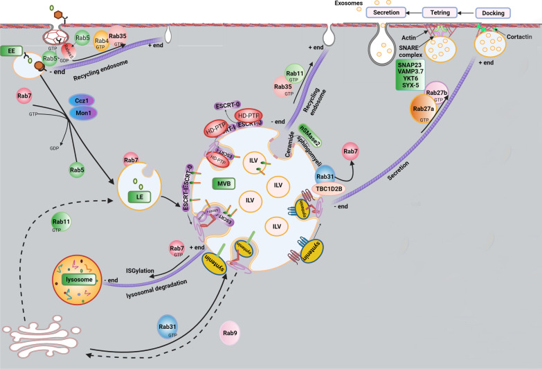
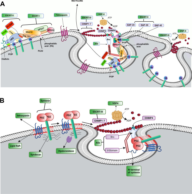
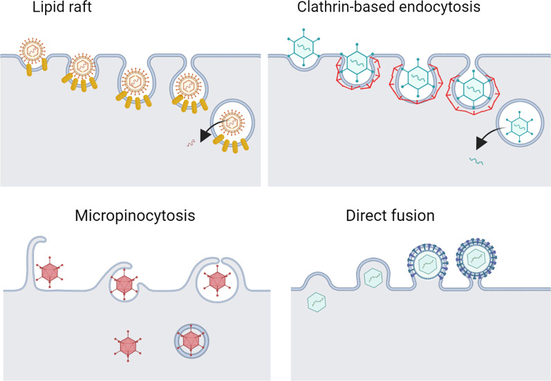
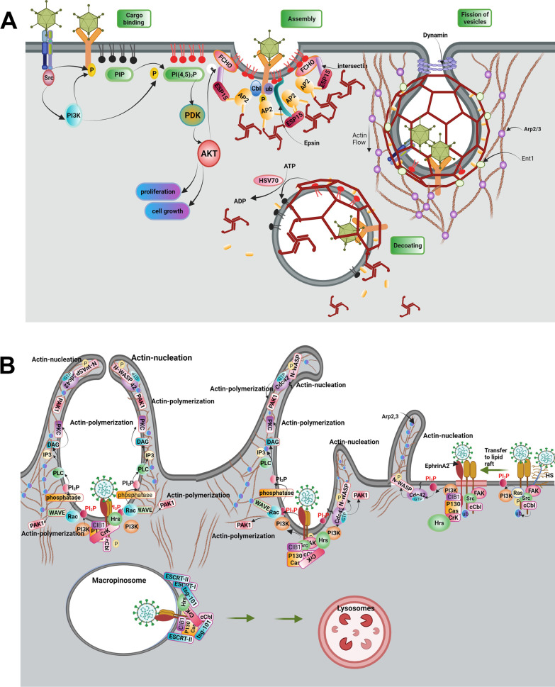
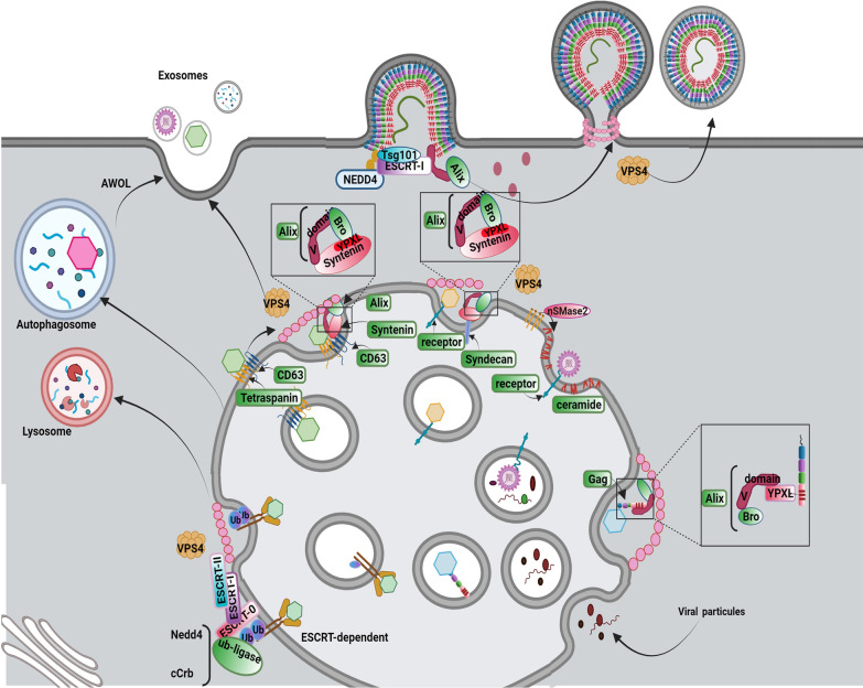
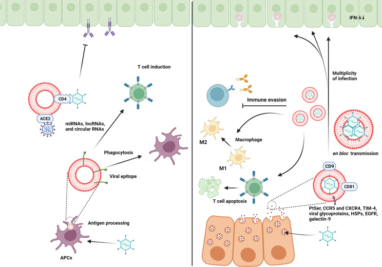
As a common belief, most viruses can egress from the host cells as single particles and transmit to uninfected cells. Emerging data have revealed en bloc viral transmission as lipid bilayer-cloaked particles via extracellular vesicles especially exosomes (Exo). The supporting membrane can be originated from multivesicular bodies during intra-luminal vesicle formation and autophagic response. Exo are nano-sized particles, ranging from 40-200 nm, with the ability to harbor several types of signaling molecules from donor to acceptor cells in a paracrine manner, resulting in the modulation of specific signaling reactions in target cells. The phenomenon of Exo biogenesis consists of multiple and complex biological steps with the participation of diverse constituents and molecular pathways. Due to similarities between Exo biogenesis and virus replication and the existence of shared pathways, it is thought that viruses can hijack the Exo biogenesis machinery to spread and evade immune cells. To this end, Exo can transmit complete virions (as single units or aggregates), separate viral components, and naked genetic materials. The current review article aims to scrutinize challenges and opportunities related to the exosomal delivery of viruses in terms of viral infections and public health. Video Abstract.

Keywords: Exosomes; Infection; Shared signaling pathways; Transmission; Viruses.


Conflict of interest statement

Authors declare there is no competing interests.

Figures

Fig. 1
Fig. 1
The generation of Exo is regulated by multiple intracellular pathways. Cargoes are sequestrated into early endosomes which are originated from endocytosis (Rab4, Rab5, Rab7, and Rab35) or Trans-Golgi network (Rab11). Exo fusion and recycling back pathways are regulated by the activity of Rab proteins (Rab 27a and Rab27b), SNARE complex (SYX-5, YKT6, VAMP3.7, SNAP23). The formation of ILVs inside the MVBs is mediated by inward invagination endosomal membrane via multiple pathways: ESCRT-dependent (ESCRT-0, I, II, II and syndecan-syntenin-ALIX axis), and ESCRT-independent pathways (ceramide-enriched microdomains and tetraspanin-enriched microdomains). Multiple pathways can distinguish the orientation of MVBs toward lysosomal depredation or fusion with the plasma membrane. ISGylation process can induce lysosomal degradation. The lysosomal degradation pathway is regulated by interaction Rab7 with Dynein and induction mobility toward microtubule minus ends. To fuse the MVBs with the membrane, various Rab-GTPases control the transport of MVBs on microtubules. Rab27 stabilizes the rearrangement of the actin cytoskeleton by improving the attachment of Cortactin, leading to MVBs docking. Docking, tethering, and releasing are three steps. The activity of SNAREs mediates the fusion of the MVB membrane with the plasma membrane
Fig. 2
Fig. 2
Exo biogenesis via conventional ESCRT-dependent pathway (A). Conventional ESCRT-dependent strategy can lead sorting of four ubiquitinated cargos into ILVs. ESCRT machinery is composed of ESCRT-0, -I, -II, and -III. The recruitment of ESCRT-0 occurs on the cytoplasmic side of the endosomal membrane. ESCRT-0 complex consists of HRS and STAM subunits to recognize the ubiquitinated cargoes. The recruitment of ESCRT-0 is triggered via the interaction of the ESCRT-0 HRS subunit with PIP3 on the endosomal membrane. The interaction of ESCRT-0 HRS with clathrin proteins enhances the clustering of ESCRT-0 to endosomal membrane microdomains. To select exosomal cargo, ESCRT-0 provides a platform for the attachment of ESCRT-I. ESCRT-I consists of four subunits: TSG-101, Mvb12, VPS37, and VPS28. Interaction between ESCRT-0 HRS subunit with ESCRT-1 TSG-101 subunit leads to physical attachment of ESCRT-0 and ESCRT-1 on endosome membrane. ESCRT-0 and -I proteins provide a binding site for of ESRT-II complex (EAP45, EAP30, and two EAP20). The physical connection is done via the ESCRT-I VPS28 subunit and ESCRT-II EAP45 subunit. Interaction of ESCRT-I with ESCRT-II can induce invagination of the endosomal membrane. The complex of ESCRT-0, -I, and -II can induce the assembly and polymerization of ESCRT-III subunits (CHMP-1, -2, -3, -4, -5, -6, and -7). Interaction of EAP20 of ESCRT-II with CHMP6 of ESCRT-III promotes the recruitment of ESCRT-III subunits. The activation of ESCRT-III promotes a chain around the neck of intraluminal vesicles. Endosomal membrane curvature is stimulated by the interaction of Alix with Mvb12 and CHMP4 subunits of ESCRT-I and ESCRT-III, respectively. Upon ILV scission, Vps4-ATPase induces ESCRT-II subunits disassociation. Role of Syndecan-Syntenin-Alix in Exo biogenesis (B). Syndecan-Syntenin-Alix stimulates Alix-ESCRT-mediated sorting in Exo. Heparanase induces Syndecans clustering and facilitates its attachment to adaptor protein Syntenin via PDZ domains. Syntenin N-terminus connects to Alix via the direct interaction with V-domain. In the end, VSP4 is recruited by the ESCRT-III complex and leads a session of ILVs into MVB
Fig. 3
Fig. 3
RAB31 inhibits the lysosomal degradation of MVBs (A). The existence of a tyrosine kinase receptor namely EGFR on the late endosomal surface activates Rab31 via tyrosine phosphorylation. The phosphorylated Rab31 interacts with flotillin proteins. Rab31-flotillin acts as a scaffold heterodimer protein and forms a budding platform for sorting EGFR and other proteins into MVB lumen in collaboration with lipid rafts. On the other hand, Rab31 recruits TBC1D2B, and the Rab31-TBC1D2B complex inactivates Rab7, preventing the lysosomal degradation of MVBs. Besides the Syntenin-Alix-ESCRT-III, Rab31-flotillin complex acts as parallel sorting machinery that drives different cargoes such as CD63, CD81, and CD9 into exosomal pathways. CD63 is a membrane protein that can interact with PDZ domains of Syntenin. Interaction N-terminal of Syntenin with the V-domain of Alix induces the recruitment of ESCRT-III by the Bro domain of Alix and the formation of ILVs. HD-PTP acts as a scaffold for the binding of ESCRT subsets during the trafficking of ubiquitinated cargo into the MVB lumen (B). HRS and STAM are subunits of ESCRT-0 and can bind to the ubiquitinated cargos on the endosomal membrane. STAM is composed of STAM (GAT) and SH3 domains. STAM domain interacts with the Bro1 domain of HD-PTP and the SH3 domain is linked to the PPRPTAPKP motif in the PTP domain of HD-PTP. ESCRT-0 is dissociated from HD-PTP by the interaction of ESCRT-I TSG-101 with the PTAP motif of the PTP domain. Then, the ESCRT-I UBAP1 subunit can interact with the FYX2L motif in the CC region of the PTP domain. In the later phase, the interaction of ESCRT-III CHMP4 subunit with the Bro domain of HD-PTP enhances the dissociation of ESCRT-0 from HD-PTP, facilitates polymerization of ESCRT-III, and drives the ubiquitinated cargo into the MVB pathway
Fig. 4
Fig. 4
Several mechanisms used by viruses to internalize the host cells
Fig. 5
Fig. 5
Internalization of the virus using Clathrin-dependent endocytosis (A). Virus attachment to a wide variety of cell receptors can induce activation and autophosphorylation of Src. Interaction of Src with cell surface receptors drives phosphorylation of receptors and activation of the PI3K/Akt/mTOR signaling pathway. PI3-K via activation of cCbl (E3ubiquitin ligase) multi-ubiquitylates EGFR, which drives phosphorylation and ubiquitination of receptor, then translocates virus bonded receptors into lipid raft. Ubiquitinated receptor drives phosphorylation and monoubiquitylation of EPS15 and monoubiquitylation of epsin. Epsin is a scaffold protein and binds to PI(4,5)2P in lipid rafts, cargo receptors, Esp15, AP2 protein, and clathrin molecules. Furthermore, the interaction of ESP15 with FCHO and intersectin proteins forms a trimeric FCHo/Eps15/ intersectin complex that can interact with AP2. Thus, AP2 is recruited for attachment to the membrane receptors and Clathrin molecules via interaction with FCHo/Eps15/ intersectin complex and Epsin proteins. Nucleation of AP2 can increase the local concentration of PIP2 by incrementing the activity of PIP kinase. Membrane fission of clathrin-coated vesicles is followed by the formation of dynamin helical oligomers at the neck of vesicles. After scission, vesicles are uncoated by the activity of the HSC70 chaperone, which dephosphorylates PI(4,5)P2 to PI4P, at the results clathrin coat is disassembled. Internalization mechanism of the virus by micropinocytosis (B). entry of the virus into cells is mediated by the initial attachment of the virus to the heparan sulfate and subsequently, integrins molecules, which is followed by induction of FAK, Src, Ras, and PI3K, signaling molecules. Activation of PI3-K recruits Ub-ligase cCbl which, both monoubiquitinates integrins molecules and leads them into the lipid raft on PM. In lipid rafts virus attached to the ubiquitinated integrins interacts with Ephrin A2 receptor; which results in the recruitment of CIB1 (adaptor protein)-p130Cas (scaffold protein)-Crk (effector protein) molecules to Ephrin A2-integrin -virus complex. CIB1-p130Cas-Crk complex recruits Hrs of ESCRT-0 to the site of macropinocytosis on the PM. Besides activated signaling of Ras and PI3-K induce actin nucleation and polymerization via activation of Cdc42-WASP, Rac1-WAVE complexes, and PAK1. Besides, PIP3 on the PM recruits phospholipase C (PLC), which leads producing of IP3 and diacylglycerol (DAG). DAG, in turn, recruits protein kinase C (PKC) to further promote actin polymerization. After the scission of macropinosomes by CtBP1and/or dynamin (is not shown here) recruitment of Tsg101 by Ephrin A2, c-Cbl, and associated signal molecules, and sequential recruitment of ESCRT I-III complex proteins on the endosome directs macropinosomes to the lysosomal degradation
Fig. 6
Fig. 6
Internalization of the virus using Caveolae-dependent endocytosis (A). Attachment of virions to the membrane receptors in membrane microdomains enriched in cholesterol and PtdSer activates receptor tyrosine kinase-like orphan receptor 1 (ROR1) and subsequently directs activation of Src, PI3K, RhoA, ROCK, CFL1 signaling pathway, at the end leads phosphorylation of Caveolin1. ROR1, as a scaffold protein, facilitates the interaction of cavin-1 and phosphorylated Caveolin1 at the PM. Also, Caveolin1 recruits EHD1,2 and Dynamin2 via binding to the Pacsin2, thus leading to the formation of the caveolae neck and session of caveolae vesicles. On the other hand, phosphorylated Caveolin1 leads to actin polymerization via the activation of Rac1, PAK1, and CFL1 signaling pathways. Caveolin1 through binding to Filamin A is associated with actin proteins and regulates caveolae vesicle trafficking. Possible mechanism of virus entry mediated by flotillin-dependent endocytosis (B). Initial attachment of the virus to the cell receptor is followed by activation of PI3K, which phosphorylates Fyn kinase, activated Fyn kinase regulates both activation of zDHHC5 and phosphorylation of Flotillins. zDHHC5 via palmitoylation of flotillin1, 2 facilitates their binding to PM. Also, phosphorylated Flotillins induce homo or heterodimerization of flotillins and the formation of invagination vesicles. Besides, activated PI3K leads to actin polymerization and changes the dynamic of the actin cytoskeleton, thus the formation of endocytic vesicles
Fig. 7
Fig. 7
Hijacking mechanisms Cellular adaptor proteins and complexes for promoting viral infection. There are several budding systems for the release of virions. Different enveloped viruses are driven toward PM and hijack the ESCRT pathway via mimic of interactions between cellular adaptors and ESCRT factors. Furthermore, virions invaginate into the MVB and are led to lysosomal degradation or hijack exosomal and autophagosomal pathways. Budding into the MVB is mediated by the hijacking of the ESCRT pathway via attachment to the ubiquitinated viral receptors and finally lysosomal degradation. Also, the entry of virions into the secretion pathway is mediated by the attachment of virions to CD63 and recruitment of the CD63-Syntenin-Alix pathway. In addition to the binding of the virus to the host endosomal receptors and lead formation of ILV by Syndecan-Syntenin-Alix, and formation of ceramide by nSMnase induce. Besides, capsid proteins of virion recruit Alix to the PM and lead assembly of ESCRT-III and formation of virion vesicles. Hijacking Alix protein can promote viral ILV budding and exosomal pathway. ILVs containing LC3 form autophagosome-like vesicles which mediate the exit of the virus through autophagosomal vesicles or AWOL
Fig. 8
Fig. 8
Opposing effects of Exo on viral infectivity and immunity

Similar articles

Cited by

References

    1. Rezabakhsh A, Sokullu E, Rahbarghazi R. Applications, challenges and prospects of mesenchymal stem cell exosomes in regenerative medicine. Stem Cell Res Ther. 2021;12(1):1–8. doi: 10.1186/s13287-021-02596-z. - DOI - PMC - PubMed
    1. Chan M-H, Chang Z-X, Huang C-YF, Lee LJ, Liu R-S, Hsiao M. Integrated therapy platform of exosomal system: hybrid inorganic/organic nanoparticles with exosomes for cancer treatment. Nanoscale Horizons; 2022. - PubMed
    1. Amini H, Rezabakhsh A, Heidarzadeh M, Hassanpour M, Hashemzadeh S, Ghaderi S, Sokullu E, Rahbarghazi R, Reiter RJ. An examination of the putative role of melatonin in exosome biogenesis. Front Cell Dev Biol. 2021;9:686551. doi: 10.3389/fcell.2021.686551. - DOI - PMC - PubMed
    1. Gurunathan S, Kang M-H, Kim J-H. A comprehensive review on factors influences biogenesis, functions, therapeutic and clinical implications of exosomes. Int J Nanomed. 2021;16:1281. doi: 10.2147/IJN.S291956. - DOI - PMC - PubMed
    1. Chaudhari P, Ghate V, Nampoothiri M, Lewis S. Multifunctional role of exosomes in viral diseases: From transmission to diagnosis and therapy. Cell Signal. 2022;110325. - PMC - PubMed