Review
. 2023 Jan 23;23(1):10.
doi: 10.1186/s40644-023-00526-1.

Contrast enhanced mammography: focus on frequently encountered benign and malignant diagnoses

Affiliations
Review

Contrast enhanced mammography: focus on frequently encountered benign and malignant diagnoses

Mindy L Yang et al. Cancer Imaging. .

Abstract

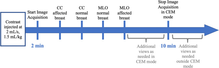
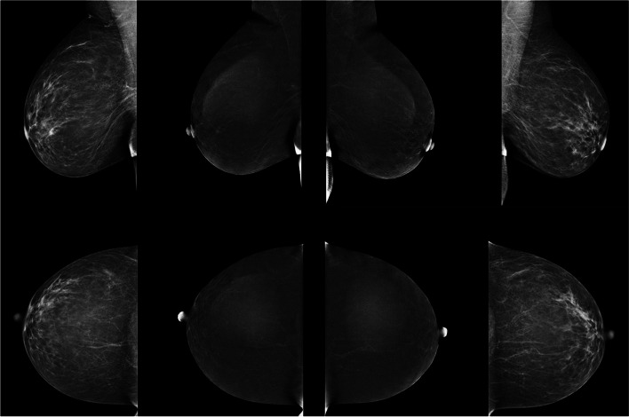
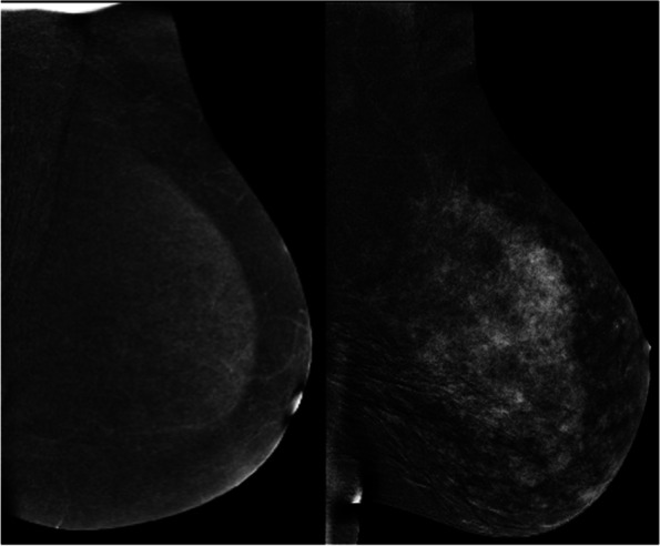
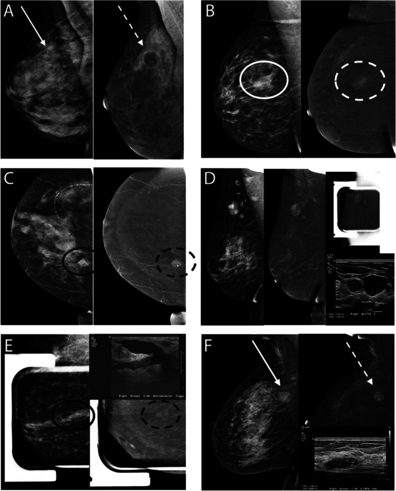
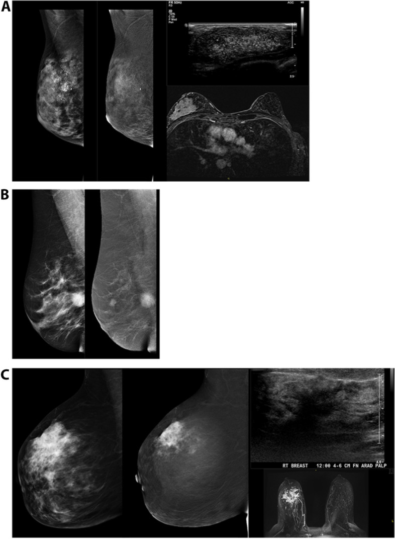
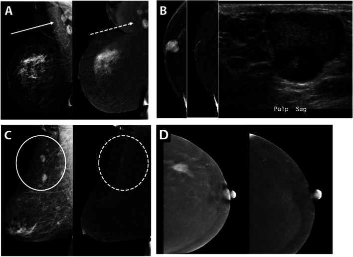
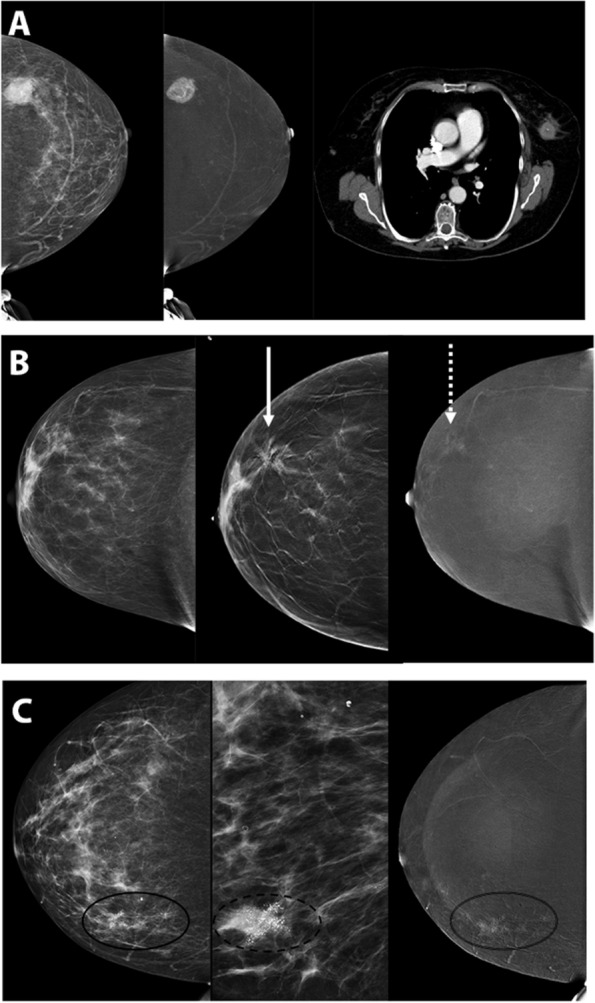
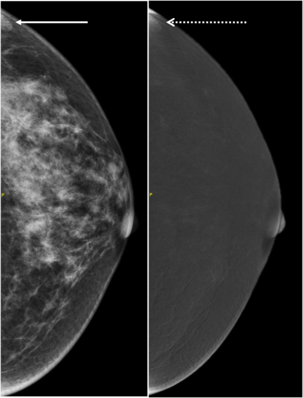
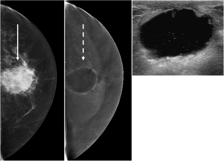
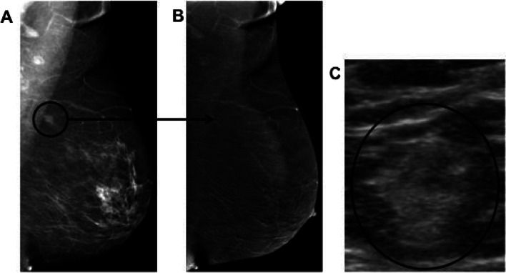
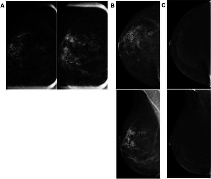
Contrast-enhanced mammography (CEM) is becoming a widely adopted modality in breast imaging over the past few decades and exponentially so over the last few years, with strong evidence of high diagnostic performance in cancer detection. Evidence is also growing indicating comparative performance of CEM to MRI in sensitivity with fewer false positive rates. As application of CEM ranges from potential use in screening dense breast populations to staging of known breast malignancy, increased familiarity with the modality and its implementation, and disease processes encountered becomes of great clinical significance. This review emphasizes expected normal findings on CEM followed by a focus on examples of the commonly encountered benign and malignant pathologies on CEM.

Keywords: Contrast mammography; Contrast-enhanced mammography; Mammography.


Conflict of interest statement

There are no potential competing interests for the authors.

The Authors declare no Competing Financial or Non-Financial Interests.

Figures

Fig. 1
Fig. 1
Current CEM protocol
Fig. 2
Fig. 2
Representative normal CEM study with standard 8 views showing the low energy (LE) and recombined images (RI) in a patient with no abnormal enhancement in either breast
Fig. 3
Fig. 3
Contrasting examples of a patient with scattered fibroglandular breast tissue who exhibits minimal BPE on RI (left) versus a patient with extremely dense breast tissue who exhibits marked BPE on RI (right)
Fig. 4
Fig. 4
Examples of Common Benign Pathologies seen on CEM. A Typical CEM appearance of cyst on LE (solid white arrow) and RI CEM (dash white arrow) with negative internal enhancement and thin rim enhancement also known as the “eclipse” sign. B Region of non-mass enhancement (dash circle) with irregular margins on LE (solid circle) corresponds to biopsy proven localized fibrocystic change. C Call back from screening mammogram for further characterization of mass in the medial breast. A 1.9 cm lobulated mass with associated solitary coarse calcification (solid black circle) demonstrates homogeneous enhancement (dash black circle) and corresponds to biopsy proven fibroepithelial lesion compatible with fibroadenoma. D Call back from screening for enlarging right axillary lymph nodes on screening mammography. CEM was performed showing enhancing circumscribed masses in the right axilla. Ultrasound revealed abnormal lymph nodes with thickened cortices. Biopsy subsequently revealed reactive changes within a biopsied lymph node. E Abnormal findings on the baseline screening mammogram, CEM recommended for further evaluation. Spot compression of retroareolar focal asymmetry (solid black circle) reveals focus of enhancement (dash black circle) at the end of the linear negative enhancement, representing a dilated duct. Corresponding ultrasound revealed intraductal hypoechoic mass, subsequently biopsy proven as intraductal papilloma. F Call back from screening mammography for bilateral abnormalities. Round mass with indistinct borders in upper inner right breast, posterior depth (solid white arrow) on LE images demonstrates enhancement on RI CEM (dash white arrow). Ultrasound of the right breast at 1:00, 8 cm from nipple, revealed an oval parallel hypoechoic circumscribed mass, subsequently biopsy proven as PASH
Fig. 5
Fig. 5
Malignant Pathologies (DCIS, IDC, ILC). A) 42-year-old woman presented with new palpable lump. Extensive pleomorphic microcalcifications throughout upper outer breast span at least 8 cm on LE images with non-mass enhancement in region of microcalcifications on RI. Ultrasound performed in this area revealed hypoechoic mass with numerous echogenic foci, subsequently biopsied revealing DCIS. A preoperative MRI revealed a large area of non-mass enhancement in the same distribution. Upon mastectomy, the pathology was upgraded to invasive ductal carcinoma. B 47-year-old woman called back from screening for new mass. Round spiculated 2.1 cm mass in the inferior central breast at posterior depth on LE images demonstrates avid enhancement on RI in addition to demonstrating smaller satellite masses in the upper central breast. After subsequent biopsy and mastectomy, findings on pathology were compatible with multicentric invasive ductal carcinoma. C 44-year-old woman presented with palpable lump in R breast. On CEM, a large high density spiculated mass at 12:00 in the mid breast on LE images is associated with avid enhancement on RI; US confirmed spiculated shadowing mass. MRI showed findings similar to CEM. Final diagnosis: invasive lobular carcinoma with associated LCIS
Fig. 6
Fig. 6
Associated Features of Malignancy and Other Entities. A Patient with multicentric breast cancer with enhancing metastatic disease to the right axilla (solid arrow corresponding to axillary lymph nodes on LE, dash arrow corresponding to enhancing axillary lymph nodes on RI). B 61-year-old man presented with palpable enhancing subareolar mass. Ultrasound correlate revealed a circumscribed hypoechoic mass with posterior enhancement, subsequently biopsied to reveal invasive ductal carcinoma with papillary features, for which a mastectomy was performed with curative intent. C 59-year-old woman with history of lymphoma presented with multiple oval enhancing masses in the breast (solid circle on LE, dash circle on RI) compatible with biopsy proven diffuse B cell lymphoma. D 56-year-old woman with known infiltrating ductal carcinoma, shown before and after neoadjuvant chemotherapy RI CEM pre-treatment image shows an enhancing spiculated mass with resolution of enhancement and mass on post-treatment RI (clip indicates an area of biopsy-proven malignancy)
Fig. 7
Fig. 7
High Risk Lesions or Borderline. A 76-year-old woman presented with new hyperdense mass in left upper outer breast on LE images, with avid enhancement and central necrosis on RI CEM. Patient unable to undergo MRI due to pacemaker but subsequent staging CT chest image shows a corresponding mass. Patient underwent left mastectomy which revealed malignant phyllodes tumor. B 40-year-old woman presenting for baseline screening mammogram, called back for architectural distortion. On CEM diagnostic workup, there is persistence of the architectural distortion (solid arrow) on LE images with minimal central enhancement (dash arrow). Ultrasound (not pictured) revealed a subtle area of distortion. Subsequent biopsy was compatible with radial scar, atypical ductal hyperplasia, intraductal papilloma. C Patient presented with suspicious microcalcifications (solid circle on LE image, dash circle on magnified view, and double line circle corresponding to non-mass enhancement on RI)
Fig. 8
Fig. 8
58-year-old woman presented for diagnostic work up for abnormal findings in both breasts. LE image shows an asymmetry (solid arrow) in the posterior far lateral left breast with corresponding enhancement on the RI (dash arrow). Upon physical exam at the time of ultrasound, this area of enhancement correlated to a skin lesion, seborrheic keratosis
Fig. 9
Fig. 9
63-year-old woman with left breast palpable abnormality 7 months post excisional biopsy for atypia. CEM shows a dense round mass (solid arrow) on LE image in the central left breast with associated rim enhancement on RI (dash arrow). On subsequent ultrasound, a fluid collection with mildly thickened wall in the area of previous excision corresponds to the mammographic finding, supporting benign etiology of postoperative seroma
Fig. 10
Fig. 10
Bilateral CEM RI in CC projections demonstrate contrast within breast vessels (white arrows) supporting adequate contrast bolus
Fig. 11
Fig. 11
Example of lack of enhancement due to small size. Patient was referred from an outside institution for abnormal screening mammogram. A CEM shows a 1.1 cm spiculated mass (black circle) on the LE image in the posterior left upper breast, axillary tail region, only seen on the MLO view. B No enhancement is seen on RI. C Subsequent ultrasound revealed an indistinct 1.2 cm echogenic mass (black circle) which correlated with the mammogram finding. Ultrasound guided biopsy confirmed invasive ductal carcinoma
Fig. 12
Fig. 12
64-year-old female was called back from screening. (A) Spot magnification views in the CC and ML projections show two groups of suspicious microcalcifications (black circles), one of which is associated with a vague focal asymmetry which persists on spot compression (B); same day CEM study again shows the two groups of microcalcifications (black circles) on the LE images (C) but with no suspicious enhancement on RI. A stereotactic needle biopsy was performed confirming low grade DCIS. This case illustrates how CEM can protect sensitivity in the example of DCIS on LE mammogram by identifying the morphologic abnormality of microcalcifications before assessing enhancement on subtraction mammogram of CEM (no enhancement is identified in this case)

Similar articles

Cited by

References

    1. Lewin JM, Isaacs PK, Vance V, Larke FJ. Dual-energy contrast-enhanced digital subtraction mammography: feasibility. Radiology. 2003;229:261–268. doi: 10.1148/radiol.2291021276. - DOI - PubMed
    1. Jm L, Patel BK, Tanna A. Contrast-enhanced mammography: a scientific review. J Breast Imaging. 2020;2(1):7–15. doi: 10.1093/jbi/wbz074. - DOI - PubMed
    1. Dromain C, Balleyguier C, Adler G, et al. Contrast-enhanced digital mammography. Eur J Radiol. 2009;69(1):34–42. doi: 10.1016/j.ejrad.2008.07.035. - DOI - PubMed
    1. Travieso-Aja MD, Maldonado-Saluzzi D, Naranjo-Santana P, et al. Diagnostic performance of contrast-enhanced dual-energy spectral mammography (CESM): a retrospective study involving 644 breast lesions. Radiol Med. 2019;124(10):1006–1017. doi: 10.1007/s11547-019-01056-2. - DOI - PubMed
    1. Dromain C, Thibault F, Diekmann F, et al. Dual-energy contrast-enhanced digital mammography: initial clinical results of a multireader, multicase study. Breast Cancer Res. 2012;14(3):R94. doi: 10.1186/bcr3210. - DOI - PMC - PubMed