

Dissolvable alginate hydrogel-based biofilm microreactors for antibiotic susceptibility assays
- PMID: 36691521
- PMCID: PMC9860113
- DOI: 10.1016/j.bioflm.2022.100103
Dissolvable alginate hydrogel-based biofilm microreactors for antibiotic susceptibility assays
Abstract
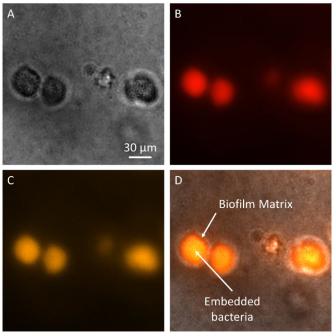
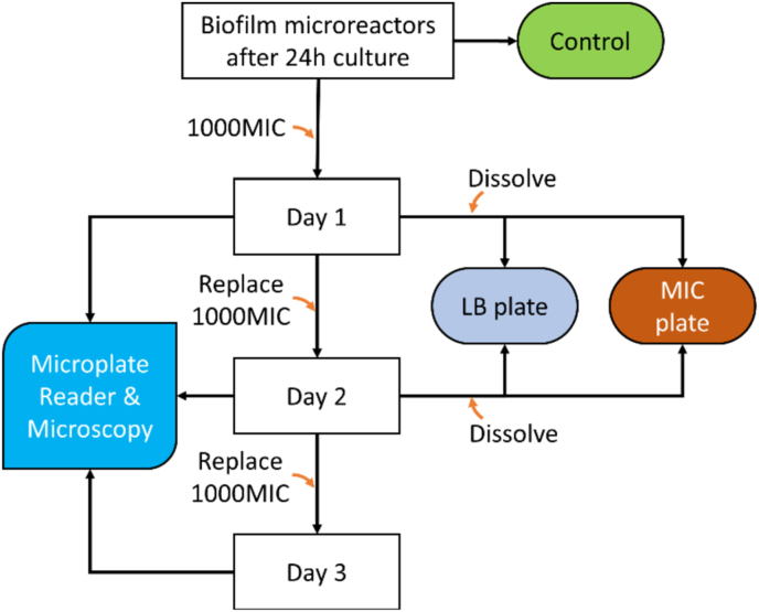
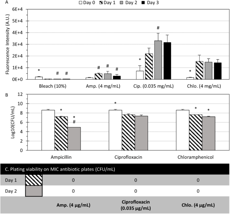
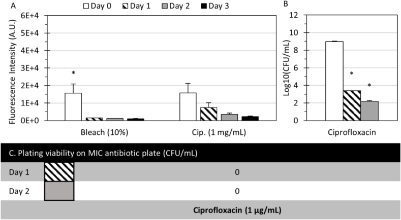
Biofilms are found in many infections in the forms of surface-adhering aggregates on medical devices, small clumps in tissues, or even in synovial fluid. Although antibiotic resistance genes are studied and monitored in the clinic, the structural and phenotypic changes that take place in biofilms can also lead to significant changes in how bacteria respond to antibiotics. Therefore, it is important to better understand the relationship between biofilm phenotypes and resistance and develop approaches that are compatible with clinical testing. Current methods for studying antimicrobial susceptibility are mostly planktonic or planar biofilm reactors. In this work, we develop a new type of biofilm reactor-three-dimensional (3D) microreactors-to recreate biofilms in a microenvironment that better mimics those in vivo where bacteria tend to form surface-independent biofilms in living tissues. The microreactors are formed on microplates, treated with antibiotics of 1000 times of the corresponding minimal inhibitory concentrations (1000 × MIC), and monitored spectroscopically with a microplate reader in a high-throughput manner. The hydrogels are dissolvable on demand without the need for manual scraping, thus enabling measurements of phenotypic changes. Bacteria inside the biofilm microreactors are found to survive exposure to 1000 × MIC of antibiotics, and subsequent comparison with plating results reveals no antibiotic resistance-associated phenotypes. The presented microreactor offers an attractive platform to study the tolerance and antibiotic resistance of surface-independent biofilms such as those found in tissues.
Keywords: Antibiotic resistance; Antibiotic susceptibility assays; Biofilm microreactors; Biofilm phenotype; Hydrogel.
© 2023 The Authors.
Conflict of interest statement
The authors declare the following financial interests/personal relationships which may be considered as potential competing interests: Xiaolong Luo and John S. Choy report financial support was provided by 10.13039/100000002National Institutes of Health. Le Hoang Phu Pham, Xiaolong Luo and Kenneth Scott Phillips have patent pending.
Figures


Similar articles
-
Effects of antibiotic treatment and phagocyte infiltration on development of Pseudomonas aeruginosa biofilm-Insights from the application of a novel PF hydrogel model in vitro and in vivo.Front Cell Infect Microbiol. 2022 Jul 26;12:826450. doi: 10.3389/fcimb.2022.826450. eCollection 2022. Front Cell Infect Microbiol. 2022. PMID: 35959369 Free PMC article.
-
Biofilm and Planktonic Antibiotic Resistance in Patients With Acute Exacerbation of Chronic Rhinosinusitis.Front Cell Infect Microbiol. 2022 Jan 17;11:813076. doi: 10.3389/fcimb.2021.813076. eCollection 2021. Front Cell Infect Microbiol. 2022. PMID: 35111699 Free PMC article.
-
Antibiotic Minimal Selective Concentrations and Fitness Costs during Biofilm and Planktonic Growth.mBio. 2022 Jun 28;13(3):e0144722. doi: 10.1128/mbio.01447-22. Epub 2022 Jun 13. mBio. 2022. PMID: 35695458 Free PMC article.
-
In Vitro Antimicrobial Susceptibility Testing of Biofilm-Growing Bacteria: Current and Emerging Methods.Adv Exp Med Biol. 2022;1369:33-51. doi: 10.1007/5584_2021_641. Adv Exp Med Biol. 2022. PMID: 33963526 Review.
-
Antimicrobial susceptibility testing in biofilm-growing bacteria.Clin Microbiol Infect. 2014 Oct;20(10):981-90. doi: 10.1111/1469-0691.12651. Epub 2014 Jun 14. Clin Microbiol Infect. 2014. PMID: 24766583 Review.
Cited by
-
Experimental Advances in the Real-Time Recording of Cross-Linking Alginate In Situ Gelation: A Review.Polymers (Basel). 2023 Jun 29;15(13):2875. doi: 10.3390/polym15132875. Polymers (Basel). 2023. PMID: 37447520 Free PMC article. Review.
-
Biofilm antimicrobial susceptibility testing: where are we and where could we be going?Clin Microbiol Rev. 2023 Dec 20;36(4):e0002423. doi: 10.1128/cmr.00024-23. Epub 2023 Oct 9. Clin Microbiol Rev. 2023. PMID: 37812003 Free PMC article. Review.
-
The landscape of biofilm models for phage therapy: mimicking biofilms in diabetic foot ulcers using 3D models.Front Microbiol. 2025 Feb 24;16:1553979. doi: 10.3389/fmicb.2025.1553979. eCollection 2025. Front Microbiol. 2025. PMID: 40066272 Free PMC article. Review.
-
Effect of Xylitol on Inhibition and Eradication of Pseudomonas aeruginosa PAO1 and Methicillin-Resistant Staphylococcus aureus Biofilms in an Alginate Bead Model.Curr Microbiol. 2024 Jul 16;81(9):272. doi: 10.1007/s00284-024-03799-2. Curr Microbiol. 2024. PMID: 39014046
-
Efficient Degradation of Alginate and Preparation of Alginate Oligosaccharides by a Novel Biofunctional Alginate Lyase with High Activity and Excellent Thermophilic Features.Mar Drugs. 2023 Mar 14;21(3):180. doi: 10.3390/md21030180. Mar Drugs. 2023. PMID: 36976229 Free PMC article.
References
Grants and funding
LinkOut - more resources
Full Text Sources
