. 2022 Dec 23;9(1):e12575.
doi: 10.1016/j.heliyon.2022.e12575. eCollection 2023 Jan.

Activation of NRF2 blocks HIV replication and apoptosis in macrophages

Affiliations

Activation of NRF2 blocks HIV replication and apoptosis in macrophages

Dating Han et al. Heliyon. .

Abstract

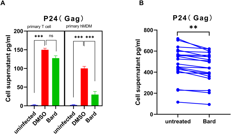
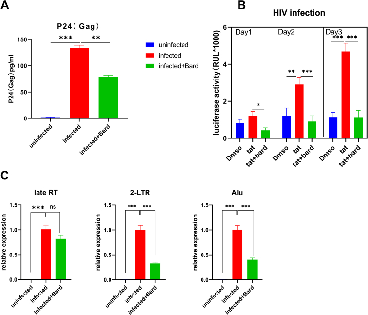
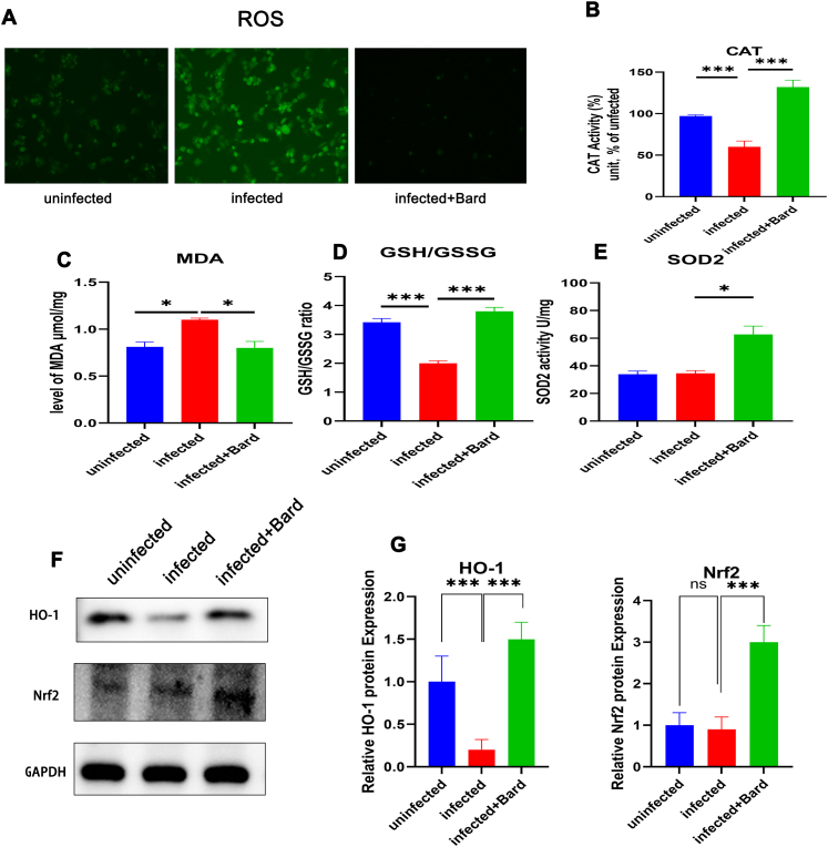
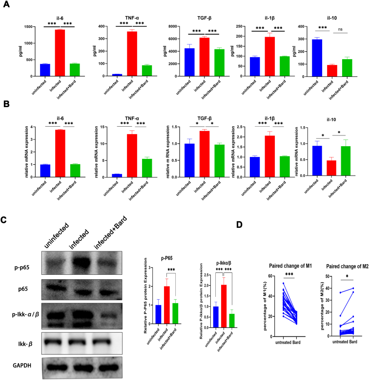
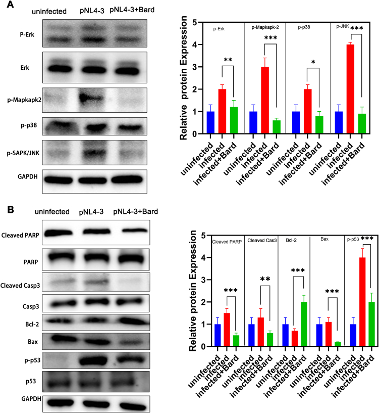
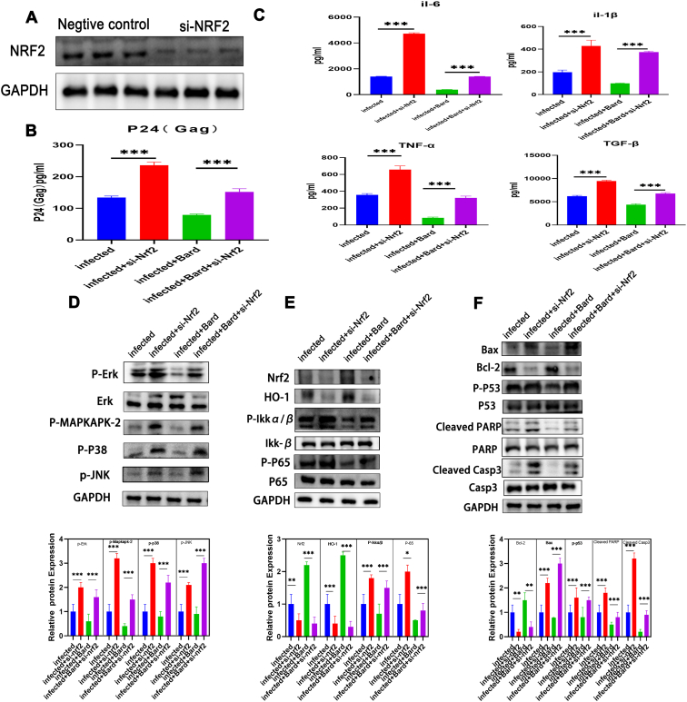
Abnormal oxidative stress caused by human immunodeficiency virus (HIV) infection affects viral replication and causes non-acquired immune deficiency syndrome-related complications in infected individuals. The transcription factor NFE2-related factor 2 (NRF2), a key regulator of oxidative stress, responds to abnormal oxidative stress by regulating the expression of NRF2-dependent cytoprotective genes. The present study aimed to determine whether inhibition of oxidative stress could control HIV replication and improve cell survival. In this study, the NRF2 activator, methyl bardoxolone, was used to treat cells for HIV infection. The effects on HIV replication and apoptosis pathways were confirmed by NRF2 activation or knockdown. The results showed that NRF2 activation could block HIV replication in macrophages before the integration phase and inhibited the expression of apoptotic pathways in virus-exposed macrophages. The study presents an unconventional anti-viral strategy of activation antioxidant response for HIV infection blocking.

Keywords: Apoptosis; HIV; Inflammation; Macrophage; Oxidative stress.


Conflict of interest statement

The authors declare no conflict of interest.

Figures

Figure 1
Figure 1
NRF2 blocks HIV in primary macrophages but not in primary T cells. (A) Primary T cells and hMDMs were either mock-infected or infected with VSV-G pseudotyped HIV-1 GFP reporter virus for 24 h. (B) Primary hMDMs from HIV-infected patients were treated with media supplemented with vehicle only (DMSO) or with 0.1 μM Bard. After 24 h of treatment, the supernatant of the cells was harvested and the level of P24(Gag) was measured using ELISA. The bar graphs represent the data for replicate experiments (n = 3). The line represents the data from the same patient (∗P < 0.05, ∗∗P < 0.01, ∗∗∗P < 0.001). NRF2, NFE2-related factor 2; hMDMs, human monocyte-derived macrophages; DMSO, dimethyl sulfoxide; Bard, methyl bardoxolone; ELISA, enzyme-linked immunosorbent assay.
Figure 2
Figure 2
NRF2 blocks HIV after entry and before or at 2-LTR circle formation. (A) PMA-induced THP-1 cells were pretreated with media supplemented with vehicle only (DMSO) or with 0.1 μM Bard for 24 h, after cells, were infected with either mock-infected or VSV-G-pseudotyped HIV-1 encoding GFP virus. After 24 h, cells were harvested for P24(Gag) detection. The bar graphs represent the data for replicate experiments (n = 3). (B) TZM-bl cells were treated with media supplemented with 100 ng/ml Tat or with 0.1 μM Bard for 24 h, 48 h, or 72 h and measured for luciferase expression, the data are presented as the mean ± SD (n = 3). (C–E) PMA-induced THP-1 cells were infected with VSV-G pseudotyped HIV-1 GFP reporter virus for 24 h after pretreated with media supplemented with vehicle only (DMSO) or with 0.1 μM Bard for 24 h. Cells were then harvested and viral DNA products were detected using real-time PCR with primer sets specific for the stage of HIV reverse transcription. (C) Relative quantities of late reverse transcription products, (D) 2-LTR circles, and (E) integrated proviruses. The bar graph represents the data for replicate experiments (n = 3). NRF2, NFE2-related factor 2; PMA, phorbol myristate acetate; GFP, green fluorescent protein; DMSO, dimethyl sulfoxide; Bard, methyl bardoxolone; 2-LTR, 2-long terminal repeat.
Figure 3
Figure 3
NRF2 activation promotes HIV induced the expression of oxides and antioxidants in macrophage cells. THP-1 cells were either mock-infected or infected with VSV-G pseudotyped HIV-1 GFP reporter virus for 24 h and treated with media supplemented with vehicle only (DMSO) or with 0.1 μM Bard. After 24 h, the levels of ROS (A) CAT(B) MDA (C) GSH/GSSG (D) SOD2 (E) in the cells were measured using assay kit and HO-1 (F) was assessed using western blotting. The bar graphs represent quantification of the Western blot band density normalized to the control for replicate experiments (n = 3). Each experiment was repeated three times with similar results. (∗P < 0.05, ∗∗P < 0.01, ∗∗∗P < 0.001). NRF2, NFE2-related factor 2; DMSO, dimethyl sulfoxide; Bard, methyl bardoxolone; ROS, reactive oxygen species; CAT, catalase; MDA, malondialdehyde; GSH, glutathione; GSSG, glutathione disulfide; SOD, superoxide dismutase; HO-1, heme oxygenase 1.
Figure 4
Figure 4
NRF2 inhibits HIV induced pro-inflammatory effects in macrophage cells. THP-1 cells were either mock-infected or infected with VSV-G pseudotyped HIV-1 GFP reporter virus for 24,h and then treated with media supplemented with vehicle only (DMSO) or with, 0.1 μM Bard. After 24 h of treatment, the supernatant and the cells were harvested and the level of cytokines was measured using ELISA (A) and cytokine mRNAs were measured using real-time PCR (B), the cellular proteins were harvested to meBiomedicineevel of phosphorylated P65 and Ikkα/β, the relative quantification of the Western blot band density was normalized to that of the control (C). The bar graphs represent the data for replicate experiments (n = 3). (D) Primary hMDMs cells from HIV-infected patients were treated with media supplemented with vehicle only (DMSO) or with 0.1 μM Bard. After 24 h of treatment, the cells were harvested for flow cytometry to measure the percentage of M1 type and M2 type macrophages; the lines represent the data from the same patient. The lines represent the data from the same patient (∗P < 0.05, ∗∗P < 0.01, ∗∗∗P < 0.001). NRF2, NFE2-related factor 2; hMDMs, human monocyte-derived macrophages; DMSO, dimethyl sulfoxide; Bard, methyl bardoxolone; ELISA, enzyme-linked immunosorbent assay.
Figure 5
Figure 5
NRF2 depress the MAPK pathway and apoptosis pathway in HIV infected macrophages cells. THP-1 cells were either mock-infected or infected withp VSV-G pseudotyped HIV-1 GFP reporter virus for 24 h and then treated with media supplemented with vehicle only (DMSO) or with 5 μM Bard. After 24 h, the levels of apoptosis pathway proteins (A) and MAPK pathway proteins (B)were assessed using western blotting. The bar graphs represent the quantification of Western blot band density normalized to that of the control for replicate experiments (n = 3) Each experiment was repeated three times with similar results. (∗P < 0.05,∗∗P < 0.01, ∗∗∗P < 0.001). NRF2, NFE2-related factor 2; MAPK, mitogen activated protein kinase; DMSO, dimethyl sulfoxide; Bard, methyl bardoxolone.
Figure 6
Figure 6
si-NRF2 rescued the function of Bard in HIV infected macrophages cells.(A) THP-1 cells were transfected with si-NRF2 or si-NC, the expression of NRF2 were measured by Western blot for the efficiency of knowndown. THP-1 cells either mock-infected or infected with VSV-G pseudotyped HIV-1 GFP reporter virus were transfected with si-NRF2 or si-NC were treated for 48 h with media supplemented with either vehicle only (DMSO) or with 0.1 μM Bard. After 24 h the supernatant of the cell was harvested and measured by ELISA for the level of P24(Gag) (B) and cytokines (C). Proteins were extracted from the cells to determine the levels of MAPK pathway proteins (C) NF-κB, NRF2, and HO-1 (D) and apoptosis pathway proteins (E) by western blotting. The bar graphs represent quantification of the Western blot band density normalized to that of the control for replicate experiments (n = 3) Each experiment was repeated three times with similar results. (∗P < 0.05, ∗∗P < 0.01, ∗∗∗P < 0.001RF2, NFE2-related factor 2; Bard, methyl bardoxolone; DMSO, dimethyl sulfoxide; ELISA, enzyme-linked immunosorbent; MAPK, mitogen activated protein kinase; HO-1, heme oxygenase 1.

Similar articles

Cited by

References

    1. Wilson E.M., Singh A., Hullsiek K.H., Gibson D., Henry W.K., Lichtenstein K., Önen N.F., Kojic E., Patel P., Brooks J.T., et al. Monocyte-activation phenotypes are associated with biomarkers of inflammation and coagulation in chronic HIV infection. J. Infect. Dis. 2014;210:1396–1406. - PMC - PubMed
    1. Carvallo L., Lopez L., Fajardo J.E., Jaureguiberry-Bravo M., Fiser A., Berman J.W. HIV-Tat regulates macrophage gene expression in the context of neuroAIDS. PLoS One. 2017;12 - PMC - PubMed
    1. Phillips D.M., Tan X., Perotti M.E., Zacharopoulos V.R. Mechanism of monocyte-macrophage-mediated transmission of HIV. AIDS Res. Hum. Retrovir. 1998;14(Suppl 1):S67–70. - PubMed
    1. Collins D.R., Lubow J., Lukic Z., Mashiba M., Collins K.L. Vpr promotes macrophage-dependent HIV-1 infection of CD4+ T lymphocytes. PLoS Pathog. 2015;11 - PMC - PubMed
    1. Ivanov A.V., Valuev-Elliston V.T., Ivanova O.N., Kochetkov S.N., Starodubova E.S., Bartosch B., Isaguliants M.G. Oxidative stress during HIV infection: mechanisms and consequences. Oxid. Med. Cell. Longev. 2016;2016 - PMC - PubMed

LinkOut - more resources