Injectable heat-sensitive nanocomposite hydrogel for regulating gene expression in the treatment of alcohol-induced osteonecrosis of the femoral head
- PMID: 36691581
- PMCID: PMC9862308
- DOI: 10.1063/5.0130711
Injectable heat-sensitive nanocomposite hydrogel for regulating gene expression in the treatment of alcohol-induced osteonecrosis of the femoral head
Abstract
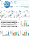
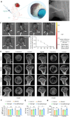
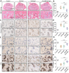
For repairing lesions, it is important to recover physiological and cellular activities. Gene therapy can restore these activities by regulating the expression of genes in lesion cells; however, in chronic diseases, such as alcohol-induced osteonecrosis of the femoral head (ONFH), gene therapy has failed to provide long-term effects. In this study, we developed a heat-sensitive nanocomposite hydrogel system with a secondary nanostructure that can regulate gene expression and achieve long-term gene regulation in lesion cells. This nanocomposite hydrogel exists in a liquid state at 25 °C and is injectable. Once injected into the body, the hydrogel can undergo solidification induced by body heat, thereby gaining the ability to be retained in the body for a prolonged time period. With the gradual degradation of the hydrogel in vivo, the internal secondary nanostructures are continuously released. These nanoparticles carry plasmids and siRNA into lesion stem cells to promote the expression of B-cell lymphoma 2 (inhibiting the apoptosis of stem cells) and inhibit the secretion of peroxisome proliferators-activated receptors γ (PPARγ, inhibiting the adipogenic differentiation of stem cells). Finally, the physiological activity of the stem cells in the ONFH area was restored and ONFH repair was promoted. In vivo experiments demonstrated that this nanocomposite hydrogel can be indwelled for a long time, thereby providing long-term treatment effects. As a result, bone reconstruction occurs in the ONFH area, thus enabling the treatment of alcohol-induced ONFH. Our nanocomposite hydrogel provides a novel treatment option for alcohol-related diseases and may serve as a useful biomaterial for other gene therapy applications.
© 2023 Author(s).
Figures


Similar articles
-
Chrysophanic acid shifts the differentiation tendency of BMSCs to prevent alcohol-induced osteonecrosis of the femoral head.Cell Prolif. 2020 Aug;53(8):e12871. doi: 10.1111/cpr.12871. Epub 2020 Jun 29. Cell Prolif. 2020. PMID: 32597546 Free PMC article.
-
Lithium chloride attenuates the abnormal osteogenic/adipogenic differentiation of bone marrow-derived mesenchymal stem cells obtained from rats with steroid-related osteonecrosis by activating the β-catenin pathway.Int J Mol Med. 2015 Nov;36(5):1264-72. doi: 10.3892/ijmm.2015.2340. Epub 2015 Sep 8. Int J Mol Med. 2015. PMID: 26352537 Free PMC article.
-
The effect of combined regulation of the expression of peroxisome proliferator-activated receptor-γ and calcitonin gene-related peptide on alcohol-induced adipogenic differentiation of bone marrow mesenchymal stem cells.Mol Cell Biochem. 2014 Jul;392(1-2):39-48. doi: 10.1007/s11010-014-2016-4. Epub 2014 Mar 15. Mol Cell Biochem. 2014. PMID: 24633961
-
The Use of Platelet-Rich Plasma for the Treatment of Osteonecrosis of the Femoral Head: A Systematic Review.Biomed Res Int. 2020 Mar 7;2020:2642439. doi: 10.1155/2020/2642439. eCollection 2020. Biomed Res Int. 2020. PMID: 32219128 Free PMC article.
-
A Mini Review: Stem Cell Therapy for Osteonecrosis of the Femoral Head and Pharmacological Aspects.Curr Pharm Des. 2019;25(10):1099-1104. doi: 10.2174/1381612825666190527092948. Curr Pharm Des. 2019. PMID: 31131747 Review.
Cited by
-
Recent advances in osteonecrosis of the femoral head: a focus on mesenchymal stem cells and adipocytes.J Transl Med. 2025 May 27;23(1):592. doi: 10.1186/s12967-025-06564-6. J Transl Med. 2025. PMID: 40426076 Free PMC article. Review.
-
Recent advances in nanomaterials for the treatment of femoral head necrosis.Hum Cell. 2024 Sep;37(5):1290-1305. doi: 10.1007/s13577-024-01102-w. Epub 2024 Jul 12. Hum Cell. 2024. PMID: 38995503 Review.
-
Positioning regulation of organelle network via Chinese microneedle.Sci Adv. 2024 Apr 19;10(16):eadl3063. doi: 10.1126/sciadv.adl3063. Epub 2024 Apr 19. Sci Adv. 2024. PMID: 38640234 Free PMC article.
-
Bioengineering strategies targeting angiogenesis: Innovative solutions for osteonecrosis of the femoral head.J Tissue Eng. 2025 Jan 24;16:20417314241310541. doi: 10.1177/20417314241310541. eCollection 2025 Jan-Dec. J Tissue Eng. 2025. PMID: 39866964 Free PMC article. Review.
-
Therapeutic effect of low-dose BMSCs-Loaded 3D microscaffold on early osteonecrosis of the femoral head.Mater Today Bio. 2024 Dec 24;30:101426. doi: 10.1016/j.mtbio.2024.101426. eCollection 2025 Feb. Mater Today Bio. 2024. PMID: 39850243 Free PMC article.
References
-
- Rogister B., Belachew S., and Moonen G., Acta Neurol. Belg. 99(1), 32–39 (1999). - PubMed
LinkOut - more resources
Full Text Sources
Research Materials

