

Rare case of extracranial chordoid meningioma adjacent to the carotid sheath: illustrative case
- PMID: 36692061
- PMCID: PMC10550698
- DOI: 10.3171/CASE22295
Rare case of extracranial chordoid meningioma adjacent to the carotid sheath: illustrative case
Abstract
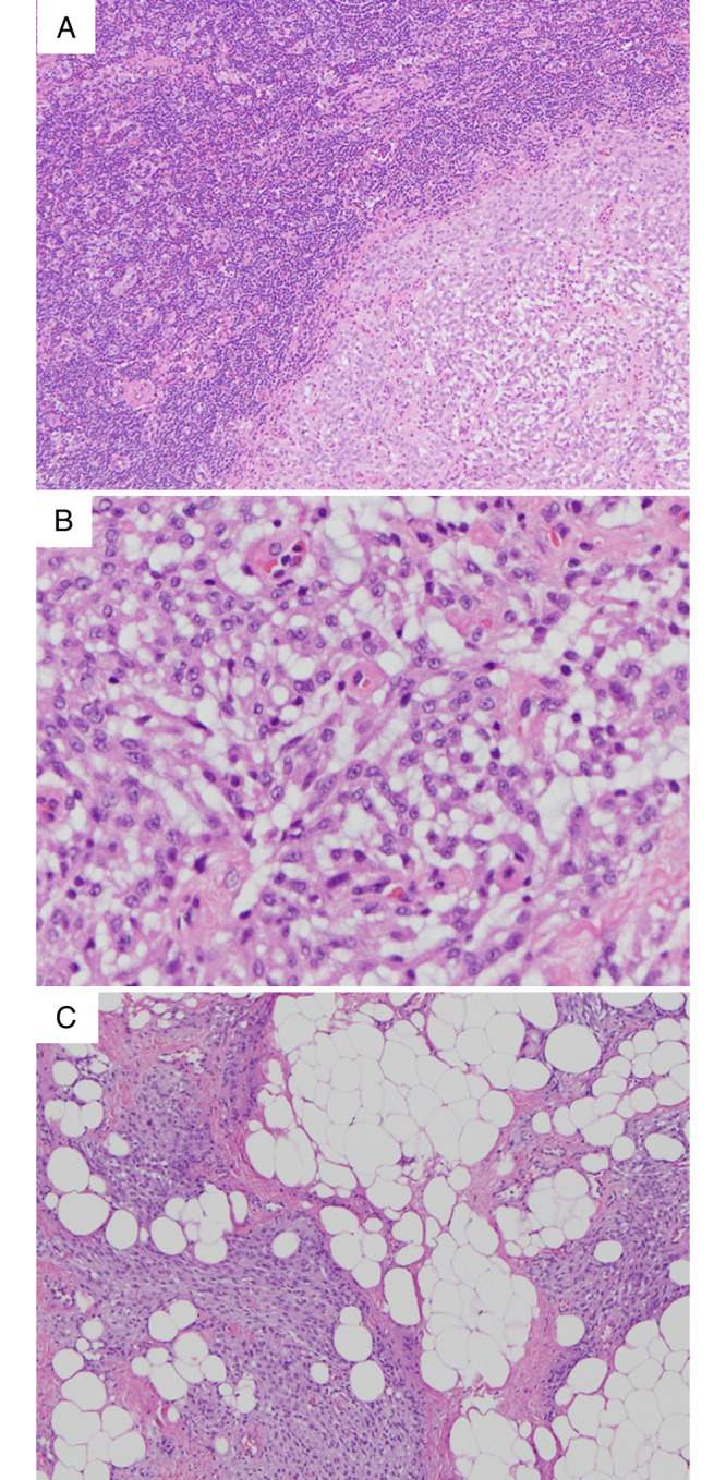
Background: Chordoid meningioma is a rare World Health Organization (WHO) grade 2 variant of meningioma with histological features resembling those of a chordoma. This tumor type is known for having an aggressive clinical course with a propensity for local recurrence. Most cases occur within the cranium, more specifically around the cerebral convexities. Although extracranial meningiomas of various subtypes have been documented, extracranial meningioma with a chordoid subtype is an extremely rare entity.
Observations: The authors herein report the case of a 51-year-old female who presented with a chief complaint of dysphagia and was found to have a neck mass abutting the carotid sheath. The patient underwent resection and final pathology results revealed a WHO grade 2 chordoid meningioma.
Lessons: This case report demonstrates an atypical case of an extracranial chordoid meningioma adjacent to the carotid sheath. To the authors' knowledge, this is the first reported case of a chordoid meningioma occurring within the soft tissue of the neck.
Keywords: carotid sheath meningioma; chordoid meningioma; extracranial chordoid meningioma; extracranial meningioma; meningioma.
Conflict of interest statement
Figures

Similar articles
-
Chordoid meningioma of the third ventricle: a case report and review of the literature.Clin Neuropathol. 2011 Mar-Apr;30(2):70-4. doi: 10.5414/npp30070. Clin Neuropathol. 2011. PMID: 21329615 Review.
-
Distinguishing chordoid meningiomas from their histologic mimics: an immunohistochemical evaluation.Am J Surg Pathol. 2009 May;33(5):669-81. doi: 10.1097/PAS.0b013e318194c566. Am J Surg Pathol. 2009. PMID: 19194275 Free PMC article.
-
Chordoid Meningioma: Differentiating a Rare World Health Organization Grade II Tumor from Other Meningioma Histologic Subtypes Using MRI.AJNR Am J Neuroradiol. 2015 Jul;36(7):1253-8. doi: 10.3174/ajnr.A4309. Epub 2015 Apr 16. AJNR Am J Neuroradiol. 2015. PMID: 25882286 Free PMC article.
-
Chordoid meningioma: rare variant of meningioma.Neuropathology. 2004 Sep;24(3):243-7. doi: 10.1111/j.1440-1789.2004.00551.x. Neuropathology. 2004. PMID: 15484703
-
Intraparenchymal chordoid meningioma: a case report and review of the literature.Int J Surg Pathol. 2012 Dec;20(6):600-5. doi: 10.1177/1066896912449043. Epub 2012 Jun 10. Int J Surg Pathol. 2012. PMID: 22689613 Review.
Cited by
-
Primary extracranial meningioma of the pelvis discovered on screening pelvic examination.BMJ Case Rep. 2023 Sep 25;16(9):e256988. doi: 10.1136/bcr-2023-256988. BMJ Case Rep. 2023. PMID: 37748813
References
-
- Burger PC, Scheithauer BW. Tumors of the Central Nervous System: Atlas of Tumor Pathology. Armed Forces Institute of Pathology; 1993. pp. 169–190. 2nd series.
-
- Surov A, Gottschling S, Bolz J, et al. Distant metastases in meningioma: an underestimated problem. J Neurooncol. 2013;112(3):323–327. - PubMed
-
- Louis DN, Perry A, Reifenberger G, et al. The 2016 World Health Organization classification of tumors of the central nervous system: a summary. Acta Neuropathol. 2016;131(6):803–820. - PubMed
-
- Couce ME, Aker FV, Scheithauer BW. Chordoid meningioma: a clinicopathologic study of 42 cases. [published correction appears in Am J Surg Pathol 2000;24(9):1316-7] Am J Surg Pathol. 2000;24(7):899–905. - PubMed
LinkOut - more resources
Full Text Sources
