. 2023 Feb;29(2):323-332.
doi: 10.3201/eid2902.220882.

Novel Prion Strain as Cause of Chronic Wasting Disease in a Moose, Finland

Novel Prion Strain as Cause of Chronic Wasting Disease in a Moose, Finland

Julianna L Sun et al. Emerg Infect Dis. 2023 Feb.

Abstract

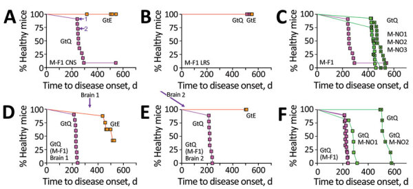
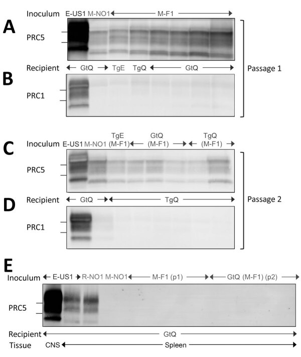
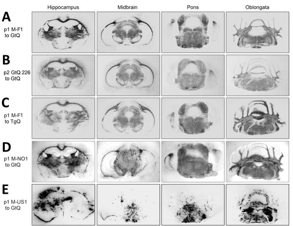
Our previous studies using gene-targeted mouse models of chronic wasting disease (CWD) demonstrated that Norway and North America cervids are infected with distinct prion strains that respond differently to naturally occurring amino acid variation at residue 226 of the prion protein. Here we performed transmissions in gene-targeted mice to investigate the properties of prions causing newly emergent CWD in moose in Finland. Although CWD prions from Finland and Norway moose had comparable responses to primary structural differences at residue 226, other distinctive criteria, including transmission kinetics, patterns of neuronal degeneration, and conformational features of prions generated in the brains of diseased mice, demonstrated that the strain properties of Finland moose CWD prions are different from those previously characterized in Norway CWD. Our findings add to a growing body of evidence for a diverse portfolio of emergent strains in Nordic countries that are etiologically distinct from the comparatively consistent strain profile of North America CWD.

Keywords: CWD; Finland; Gt; Nordic; Norway; Tg; chronic wasting disease; gene-targeted; mice; moose; prions; transgenic.


Figures

Figure 1
Figure 1
Transmission properties of Finland moose chronic wasting disease (CWD) prions in GtQ and GtE mice. Survival curves of intracerebrally inoculated GtQ and GtE mice are shown. A–C) Primary transmissions; D–F) secondary transmissions. A, B) Transmission to GtE mice (orange squares) and GtQ mice (magenta squares) of CNS homogenate from M-F1 (A) and LRS tissue homogenate from M-F1 (B). Arrows in GtQ mouse brains 1 and 2 (A) used for serial transmissions (D and E). C) Survival of GtQ mice infected with M-F1 from (A) (magenta squares) compared with Norway moose CWD isolates M-NO1 (green squares), M-NO2 (dotted green squares), and M-NO3 (crossed green squares). D) Serial passage of M-F1 prions from GtQ mouse brain 1 to GtE (orange squares) and GtQ (magenta squares). E) Serial passage of M-F1 prions from GtQ mouse brain 2 to GtE and GtQ mice. F) incubation times in GtQ mice of GtQ-passaged M-F1 (brains 1 and 2) from mice in panels D and E (magenta squares) compared with GtQ-passaged M-NO1 (green squares) and M-NO2 (dotted green squares). CNS, central nervous system; GtE, CWD-susceptible gene-targeted mice in which the prion protein coding sequence was replaced with one encoding glutamate at codon 226; GtQ, CWD-susceptible gene-targeted mice in which the prion protein coding sequence was replaced with one encoding glutamine at codon 226; LRS, lymphoreticular system; M-F1, Finland moose 1, M-NO1: Norway moose 1, M-NO2: Norway moose 2, M-NO3: Norway moose 3; p1, primary transmissions; p2, secondary transmissions.
Figure 2
Figure 2
Western blot analyses of cervid prion protein (PrP) scrapie in mice infected with Finland, Norway, and North America chronic wasting disease (CWD) isolates. A, B) Western blots of CNS homogenates from GtQ, TgQ, and TgE mice after primary transmissions E-US1, Norway moose CWD (M-NO1), or Finland moose CWD (M-F1) probed with monoclonal antibody (mAb) PRC5 (A) or PRC1 (B). C–D) Western blots of CNS homogenates of GtQ and TgQ mice infected with E-US1 isolate, Norway isolate M-NO1, and M-F1 passaged through TgE, GtQ, or TgQ, referred to as TgE (M-F1), GtQ (M-F1), and TgQ (M-F1), respectively, probed with mAb PRC5 (C) or PRC1 (D). E) Lane 1, CNS homogenate of GtQ mice infected with E-US1 isolate. Remaining lanes are spleen homogenates from GtQ mice infected with E-US1 isolate, Norway reindeer isolate R-NO1, Norway moose isolate M-NO1, and spleens from infected GtQ mice during primary and secondary transmissions of M-F1. The positions of 25 and 20 kDa molecular weight markers are shown to the left of immunoblots. CNS, central nervous system; E-US1, US elk 1; GtQ, CWD-susceptible gene-targeted mice in which the prion protein coding sequence was replaced with one encoding glutamine at codon 226; M-F1, Finland moose 1; M-NO1, Norway moose 1; PRC1, mAb PCR1; PRC5, mAb PCR5; TgE, transgenic mice expressing cervid PrP with glutamate at residue 226; TgQ, transgenic mice expressing cervid PrP with glutamine at residue 226.
Figure 3
Figure 3
Global central nervous system distribution of cervid prion protein scrapie in Tg and Gt mice infected with Finland, Norway, and North America chronic wasting disease (CWD) cryostat coronal brain sections taken at the level of the hippocampus, midbrain, pons, and oblongata, transferred to slides and then to nitrocellulose. Sections were proteinase K–treated and immunoprobed with monoclonal antibody PRC5 after denaturation. A, B) Passage 1 and 2 of M-F1 in GtQ mice. C) TgQ mice infected with M-F1. D) GtQ mice infected with Norway M-NO1 CWD. E) GtQ mice infected with CWD isolate M-US1. Gt, gene-targeted; GtQ, CWD-susceptible gene-targeted mice in which the prion protein coding sequence was replaced with one encoding glutamine at codon 226; M-F1: Finland moose 1; M-NO1, Norway moose 1; M-US1, US moose 1; p1, primary transmissions; p2, secondary transmissions; Tg, transgenic; TgQ, transgenic mice expressing cervid PrP with glutamine at residue 226.
Figure 4
Figure 4
Lesion profiling in GtQ mice infected with Finland and Norway moose chronic wasting disease (CWD) isolates. Lesion profiles in groups of GtQ mice (CWD-susceptible gene-targeted mice in which the prion protein coding sequence was replaced with one encoding glutamine at codon 226) infected with M-F1 (magenta symbols) and Norway moose isolate M-NO1 (green symbols). For M-F1, open circles and solid lines depict primary passage; dotted circles and dashed lines depict second passage. Data points represent the mean +SEM of >5 GtQ mice per group. Brain-scoring areas: medulla (1), cerebellum (2), superior colliculus (3), hypothalamus (4), thalamus (5), hippocampus (6), septum (7), retrosplenial and adjacent motor cortex (8), and cingulate and adjacent motor cortex (9). M-F1, Finland moose 1; M-NO1, Norway moose 1.
Figure 5
Figure 5
Immunohistochemical analyses of disease-associated prion protein (PrP) in the CNS of transgenic (Tg) and gene-targeted (Gt) mice infected with Finland, Norwway, and North America chronic wasting disease (CWD) isolates. A) GtQ mice (CWD-susceptible Gt mice in which the PrP coding sequence was replaced with one encoding glutamine at codon 226) infected with CWD isolate from Finland moose 1 (MF-1). B) TgE mice (Tg mice expressing cervid PrP with glutamate at residue 226) infected with M-F1. C) GtQ mice infected with CWD isolate from Norway moose 1 (M-NO1). D) GtQ mice infected with CWD isolate from US moose 1. Arrows in panels A, B, and C indicate small puncta of PrP. Immunohistochemistry sections were stained with fragment antigen binding D18. Original magnification ×10.
Figure 6
Figure 6
Responses of Finland, Norway, and North America moose chronic wasting disease (CWD) prions propagated in CWD-susceptible mice to increasing concentrations of GdnHCl. A) Responses of CerPrPSc in central nervous system (CNS) of diseased GtQ mice to proteinase K treatment after denaturation with increasing concentrations of GdnHCl. Magenta symbols indicate M-F1 CWD, green symbols M-NO1 CWD, and blue symbols M-US1 CWD. B) Denaturation profiles of cervid PrP scrapie in CNS of GtQ mice infected with M-F1. Magenta squares and solid lines depict passage 1; dotted magenta squares and dashed lines depict GtQ (M-F1) brains 1 and 2 (passage 2). C) Denaturation profiles of cervid PrP scrapie in the CNS of TgQ mice infected with M-F1 (magenta circles), TgQ mice infected with M-F1 passaged through TgE mice [TgE (M-F1)] (orange circles), and TgQ mice infected with M-F1 passaged through TgQ mice [TgQ (M-F1)] (dotted magenta circles). Proteinase K–resistant PrPSc was quantified by densitometry of immunoprobed dot blots and plotted against GdnHCl concentration. Sigmoidal dose-response curves were plotted using a 4-parameter algorithm and nonlinear least-square fit. Error bars indicate +SEM of data from analyses of >3 animals per group. GdnHCl1/2 values (mol/L) for each infection are reported on the right-hand side of each graph. Significance calculated by pairwise analysis of GdnHCl1/2 values from best fit curves. Fapp, fraction of apparent PrPSc = (maximum signal-individual signal)/(maximum signal-minimum signal); GdnHCl, guanidine hydrochloride; GdnHCl1/2, guanidine hydrochloride half-maximal denaturation; GtQ, CWD-susceptible gene-targeted mice in which the prion protein coding sequence was replaced with one encoding glutamine at codon 226; NS, not significant; PK, proteinase K; PrP, prion protein; TgE, transgenic mice expressing cervid PrP with glutamate at residue 226; TgQ, transgenic mice expressing cervid PrP with glutamine at residue 226.

Similar articles

Cited by

References

    1. Riek R, Hornemann S, Wider G, Billeter M, Glockshuber R, Wüthrich K. NMR structure of the mouse prion protein domain PrP(121-231). Nature. 1996;382:180–2. 10.1038/382180a0 - DOI - PubMed
    1. Kraus A, Hoyt F, Schwartz CL, Hansen B, Artikis E, Hughson AG, et al. High-resolution structure and strain comparison of infectious mammalian prions. Mol Cell. 2021;81:4540–4551.e6. 10.1016/j.molcel.2021.08.011 - DOI - PubMed
    1. Kang HE, Bian J, Kane SJ, Kim S, Selwyn V, Crowell J, et al. Incomplete glycosylation during prion infection unmasks a prion protein epitope that facilitates prion detection and strain discrimination. J Biol Chem. 2020;295:10420–33. 10.1074/jbc.RA120.012796 - DOI - PMC - PubMed
    1. Basler K, Oesch B, Scott M, Westaway D, Wälchli M, Groth DF, et al. Scrapie and cellular PrP isoforms are encoded by the same chromosomal gene. Cell. 1986;46:417–28. 10.1016/0092-8674(86)90662-8 - DOI - PubMed
    1. Hoyt F, Standke HG, Artikis E, Schwartz CL, Hansen B, Li K, et al. Cryo-EM structure of anchorless RML prion reveals variations in shared motifs between distinct strains. Nat Commun. 2022;13:4005. 10.1038/s41467-022-30458-6 - DOI - PMC - PubMed

LinkOut - more resources