. 2023 Mar;80(3-4):77-92.
doi: 10.1002/cm.21745. Epub 2023 Feb 8.

Acetylation of fission yeast tropomyosin does not promote differential association with cognate formins

Affiliations

Acetylation of fission yeast tropomyosin does not promote differential association with cognate formins

Qing Tang et al. Cytoskeleton (Hoboken). 2023 Mar.

Abstract

It was proposed from cellular studies that S. pombe tropomyosin Cdc8 (Tpm) segregates into two populations due to the presence or absence of an amino-terminal acetylation that specifies which formin-mediated F-actin networks it binds, but with no supporting biochemistry. To address this mechanism in vitro, we developed methods for S. pombe actin expression in Sf9 cells. We then employed 3-color TIRF microscopy using all recombinant S. pombe proteins to probe in vitro multicomponent mechanisms involving actin, acetylated and unacetylated Tpm, formins, and myosins. Acetyl-Tpm exhibits tight binding to actin in contrast to weaker binding by unacetylated Tpm. In disagreement with the differential recruitment model, Tpm showed no preferential binding to filaments assembled by the FH1-FH2-domains of two S. pombe formins, nor did Tpm binding have any bias towards the growing formin-bound actin filament barbed end. Although our in vitro findings do not support a direct formin-tropomyosin interaction, it is possible that formins bias differential tropomyosin isoform recruitment through undiscovered mechanisms. Importantly, despite a 12% sequence divergence between skeletal and S. pombe actin, S. pombe myosins Myo2 and Myo51 exhibited similar motile behavior with these two actins, validating key prior findings with these myosins that used skeletal actin.

Keywords: S. pombe; acetylation; actin; formin; myosin; tropomyosin.


Conflict of interest statement

Conflict of interest statement

The authors declare no conflict of interest.

Figures
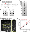
Figure 1.
Figure 1.. Expression and purification of polymerization-competent fission yeast actin from Sf9 insect cells.
(A) Schematic illustration of the actin expression construct showing the C-terminal region of S. pombe actin (amino acid 371–375), followed by a linker (zigzag line), thymosin β4, and His6. The numbering of the amino acids takes into account that Met1 is not removed in yeast actins (Cook et al., 1991). The red bold arrows indicate the preferred chymotryptic cleavage sites in both constructs, the dotted arrow indicates the less preferred cleavage site in the WT construct. (B) 3 μM S. pombe actin, obtained from chymotrypsin-cleavage of the WT actin-thymosin β4 construct, was incubated in polymerization buffer (10 mM imidazole pH 7.5, 50 mM KCl, 4 mM MgCl2, 1 mM EGTA, 1 mM DTT, and 0.2 mM MgATP) at 30°C for 3 h. The Coomassie-stained gel shows the total (T), supernatant (S), and pellet (P) fractions following high-speed centrifugation. (C) Coomassie-stained 4–12% SDS-PAGE of purified S. pombe actin(Y371H)–thymosin β4-His6 before (lane 1) and after (lane 3) cleavage and removal of the thymosin β4-His6 moiety. Lane 2, molecular mass standards. * indicates minor bands resulting from proteolysis at sites other than F375. (D) Left, A representative image showing polymerized S. pombe F-actin (Y371H) filaments observed by TIRF microscopy. Scale bar, 10 μm. Right, a time montage showing an elongating S. pombe actin filament of the zoomed region (yellow open box) from the left panel. The yellow arrowheads denote the barbed end. Scale bar, 5 μm. (E) Actin polymerization rates of chicken skeletal actin (black circles) and S. pombe actin(Y371H) (red triangles) as a function of actin concentration, as observed by TIRF microscopy using LifeAct-GFP. The assembly and disassembly rate constants and critical concentrations obtained from the linear fits to the data are given in Table 1. Buffer: 10 mM imidazole pH 7.5, 50 mM KCl, 4 mM MgCl2, 1 mM EGTA, 1 mM DTT, 0.2 mM MgATP. Temp: 25 °C. n = 25–32 filaments per condition. Two independent actin preparations were used at each actin concentration.
Figure 2.
Figure 2.. Growth characteristics of actin mutant Y371H fission yeast are similar to WT.
(A) Micrographs of WT and actin(Y371H) fission yeast cells showing morphology (DIC) and organization of actin networks (Lifeact-GFP). Scale bar, 5 μm. (B) Growth curves of WT and actin(Y371H) strains. 3 replicates of each strain are shown. Average time to half-maximum OD600 of each strain was calculated from 15 replicates each. (C) Micrographs of WT and actin-Y371H cells in DIC (morphology), and methanol fixed cells stained with DAPI (nuclei) and Calcofluor (septa). Scale bar, 5 μm.
Figure 3.
Figure 3.. Acetylation of fission yeast Tpm increases the apparent affinity for fission yeast actin and slows actin polymerization dynamics.
(A) Acetylated Tpm migrates slower than unacetylated Tpm on a charge gel. (B) Affinity of acetylated vs unacetylated S. pombe Tpm for S. pombe actin from a co-sedimentation experiment. Three experiments with two independent actin preparations were performed. Data points were fit to the Hill equation. Kapp is 2.1 × 10⁶ M−1 for acetylated Tpm, and 0.8 × 10⁶ M−1 for unacetylated Tpm. The Hill coefficient is 2.4 for acetylated Tpm, and 4.3 for unacetylated Tpm. Buffer: 10 mM imidazole, pH 7.5, 150 mM NaCl, 2 mM MgCl2, 1 mM EGTA and 1 mM DTT. (C) S. pombe actin polymerization rates in the absence or presence of acetylated Tpm (AcTpm) observed by TIRF microscopy. The critical concentrations (0.22 μM for actin, 0.13 μM for actin-AcTpm) and the depolymerization rates (4.1 subunits s−1 for actin, 1.6 subunits s−1 for actin-AcTpm) were extrapolated from the linear fits. n = 22–39 filaments per condition. Two independent actin preparations were used for each actin concentration per condition.
Fig 4.
Fig 4.. Acetylated fission yeast Tpm does not show a binding preference toward the fission yeast actin barbed end that is associated with contractile ring formin Cdc12 (FH1-FH2).
(A) Acetylated Tpm Cdc8 (D142C) migrates slower than unacetylated Tpm on a charge gel. (B) Representative kymographs of an S. pombe actin filament barbed end elongation over time observed through TIRF microscopy in the presence of 3 μM S. pombe actin, 1 nM S. pombe formin Cdc12, 3 μM S. pombe profilin Cdc3, and 0.75 μM acetylated S. pombe Tpm (D142C). The formin (SNAP-Surface-649-labeled; cyan) and Tpm D142C (TMR-labeled; magenta) are directly labeled. Actin (green) is visualized using Lifeact-GFP. Left panel, 3-color composite. Right panel, 2-color composite of the left panel (only the formin and Tpm are shown). (C) Actin (1 μM) barbed end polymerization rates in the presence of 0–4 nM of formin Cdc12 and 1 μM of profilin, with (blue) or without (red) 1 μM acetylated Tpm. Actin was visualized with Lifeact-GFP. (D) The average number of total actin filaments observed in a 54 × 54 μm2 area after 2 min observed in TIRF microscopy in conditions described in C). n = 37–41 filaments per condition. Two independent actin preparations were tested in each condition. Kolmogorov-Smirnov test was performed. * P < 0.05, **** P < 0.0001, ns, not significant.
Fig. 5.
Fig. 5.. Fission yeast actin polymerized with cable formin For3 (FH1-FH2) recruits more acetylated than unacetylated Tpm and shows no preference for binding to the actin filament end.
(A) 12 % SDS-PAGE showing SNAP-For3 (FH1-FH2)-HIS6 expressed and purified from Sf9 cells. Lane 1, protein standards. The * in lane 2 indicates a minor breakdown product that reacts with anti-HIS6 antibody. (B) Representative kymographs showing an S. pombe actin filament barbed end elongation in the presence of 1 μM S. pombe actin, 40 nM S. pombe For3, 1 μM S. pombe profilin Cdc3, and 0.25 or 2.8 μM S. pombe Tpm Cdc8 (D142C) over time. The actin (green) is visualized by using Lifeact-GFP, the Tpm D142C (TMR-labeled; magenta) is directly labeled. The For3 is unlabeled. Left panel: polymerizing actin is partially decorated by acetylated Tpm in the presence of 0.25 μM acetylated Tpm and 40 nM For3. Right panels, two examples showing very scarce regions of F-actin were bound to Tpm when 2.8 μM of unacetylated Tpm (D142C) and 40 nM For3 were present during actin polymerization, and no barbed end preference of Tpm binding to the actin filaments was observed. The open arrowheads showing the narrow, Tpm-bound regions on actin. (C) Actin (1 μM) barbed end elongation rates in the presence of 0–60 nM of formin For3 and 1 μM of profilin, with (blue triangle) or without (red circles) 1 μM acetylated Tpm. (D) The average number of total actin filaments observed in a 54 × 54 μm2 area after 2 min observed in TIRF microscopy in conditions described in C). n = 40–60 filaments per condition. Two independent actin preparations were tested in each condition. Kolmogorov-Smirnov test was performed. * P < 0.05, **** P < 0.0001, ns, not significant.
Fig 6.
Fig 6.. Myo2 moves fission yeast or skeletal actin filaments similarly in an in vitro motility assay.
(A) In vitro motility speeds of unphosphorylated fission yeast contractile ring myosin Myo2 (full length), with directly labeled (NHS-rhodamine) skeletal actin (black) and S. pombe actin (red) at 30 °C in buffer containing 50 mM KCl, 0.5 % methylcellulose, and 1 mM MgATP. Lower panel, motility speed distribution in the presence of 2 μM acetylated Tpm. n = 120–300 filaments per condition for each actin. Statistics are shown in Table 4. (B) In vitro motility speeds of phosphorylated Myo2 with directly labeled S. pombe actin with (blue triangles) or without (red circles) acetylated Tpm. The bare actin shows a wider distribution of speeds compared to actin-Tpm. n = 170–300 filaments per condition. (C) Number of S. pombe actin filaments bound to the phosphorylated (magenta) or unphosphorylated (cyan) Myo2-coated surface (128 μm × 128 μm), in the absence of methylcellulose at different KCl concentrations. Acetylated Tpm was added to 2 μM when indicated. Note that Myo2 did not bind actin filaments at 150 mM KCl in the absence of Tpm (Pollard et al., 2017). The data were collected from experiments using two independent actin preparations. Unpaired t-test with Welch’s correction, * P < 0.05, ** P < 0.01.
Fig 7.
Fig 7.. Myo51-Rng8/9 moves fission yeast or skeletal actin filaments similarly in an in vitro motility assay.
(A) In vitro motility speeds of fission yeast contractile ring and actin cable myosin Myo51-Rng8/9 (full length) with directly labeled (NHS-rhodamine) skeletal actin (black) and S. pombe actin (red) at 30 °C, in the buffers containing 50–150 mM KCl, 0.5–0.7% methylcellulose, and 1.5 mM MgATP. n = 80–130 filaments per condition for each species. Data were collected from experiments using two independent actin preparations. (B) 1 μM of Myo51 tail-Rng8/9 pellets with either S. pombe or skeletal actin in the presence of acetylated Tpm. Actin concentration, 4 μM; acetylated Tpm, 2 μM; 150 mM NaCl. The supernatant (S) and pellet (P) fractions are resolved on a 12 % SDS-PAGE.

Similar articles

Cited by

References

    1. Alioto SL, Garabedian MV, Bellavance DR, and Goode BL (2016). Tropomyosin and Profilin Cooperate to Promote Formin-Mediated Actin Nucleation and Drive Yeast Actin Cable Assembly. Current biology : CB. - PMC - PubMed
    1. Balasubramanian MK, Helfman DM, and Hemmingsen SM (1992). A new tropomyosin essential for cytokinesis in the fission yeast S. pombe. Nature 360, 84–87. - PubMed
    1. Barua B, Fagnant PM, Winkelmann DA, Trybus KM, and Hitchcock-DeGregori SE (2013). A periodic pattern of evolutionarily conserved basic and acidic residues constitutes the binding interface of actin-tropomyosin. J Biol Chem 288, 9602–9609. - PMC - PubMed
    1. Bendezú FO, and Martin SG (2011). Actin cables and the exocyst form two independent morphogenesis pathways in the fission yeast. Molecular Biology of the Cell 22, 44–53. - PMC - PubMed
    1. Bohnert KA, Grzegorzewska AP, Willet AH, Vander Kooi CW, Kovar DR, and Gould KL (2013). SIN-dependent phosphoinhibition of formin multimerization controls fission yeast cytokinesis. Genes & Development 27, 2164–2177. - PMC - PubMed

Publication types

MeSH terms