Review
. 2023 Jun;252(6):691-712.
doi: 10.1002/dvdy.567. Epub 2023 Feb 9.

The critical role of cardiolipin in metazoan differentiation, development, and maturation

Affiliations
Review

The critical role of cardiolipin in metazoan differentiation, development, and maturation

Melissa Olivar-Villanueva et al. Dev Dyn. 2023 Jun.

Abstract

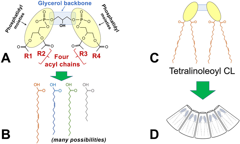
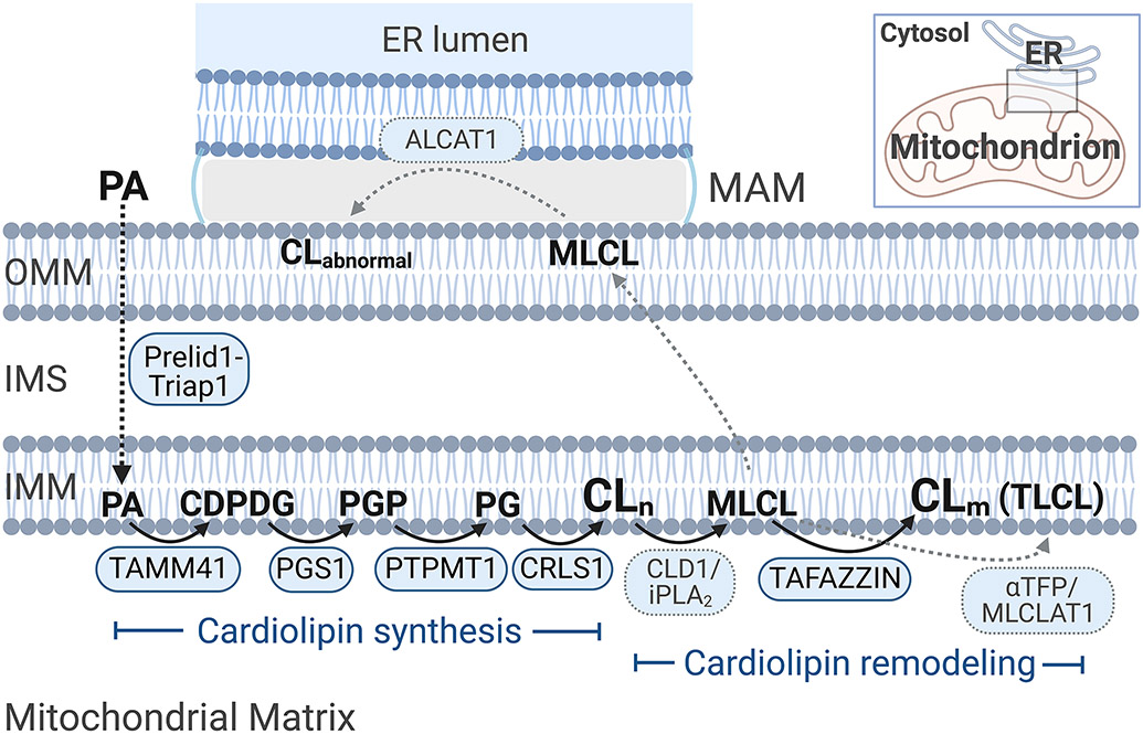
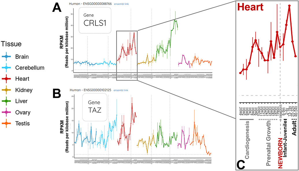
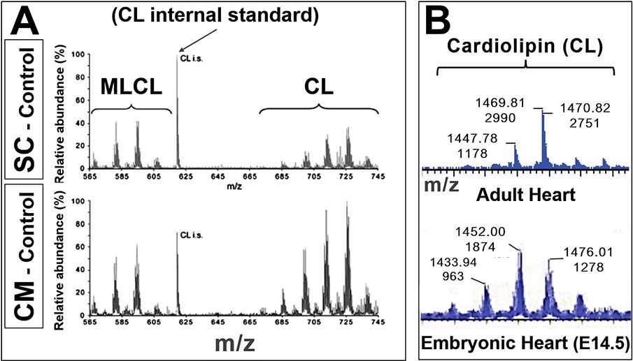
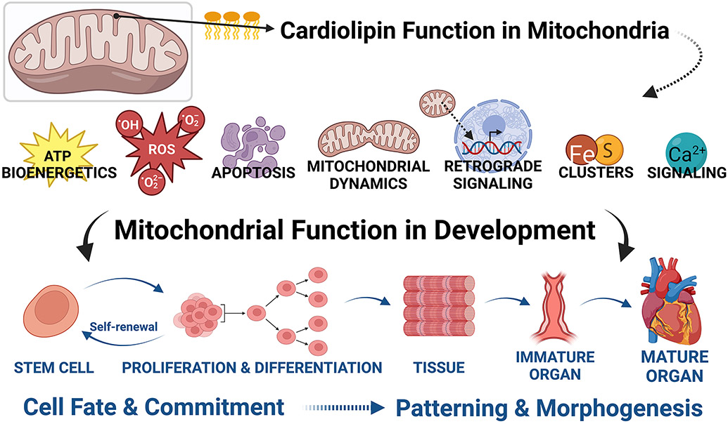
Cardiolipins are phospholipids that are central to proper mitochondrial functioning. Because mitochondria play crucial roles in differentiation, development, and maturation, we would also expect cardiolipin to play major roles in these processes. Indeed, cardiolipin has been implicated in the mechanism of three human diseases that affect young infants, implying developmental abnormalities. In this review, we will: (1) Review the biology of cardiolipin; (2) Outline the evidence for essential roles of cardiolipin during organismal development, including embryogenesis and cell maturation in vertebrate organisms; (3) Place the role(s) of cardiolipin during embryogenesis within the larger context of the roles of mitochondria in development; and (4) Suggest avenues for future research.

Keywords: Tafazzin; mitochondria; oxidative phosphorylation; phospholipids; signaling.


Conflict of interest statement

Conflict of interest disclosure: The authors declare they have no conflicts of interest to disclose.

Figures

FIGURE 1.
FIGURE 1.. Cardiolipin structure, an overview.
A) The schematic shows the key features of cardiolipin: A glycerol backbone and two phosphatidyl moieties that together form the dimeric phosphatidylglycerol head group; and 4 acyl chains (designated R1-R4). B) Each of the four acyl chains may vary by length, degrees of saturation, and positions of double bonds, resulting in an enormous possible diversity of acyl chain combinations. C) The most abundant form of cardiolipin in the mammalian heart is tetralinoleoyl cardiolipin (CL), in which the four acyl chains are linoleic acid. D) Tetralinoleoyl CL has a unique conical shape that is critical to its biophysical properties within the lipid membrane.
FIGURE 2.
FIGURE 2.. Cardiolipin biosynthetic and remodeling pathways.
Cardiolipin synthesis: Phosphatidic acid (PA) is the central precursor for the biosynthesis of glycerolipids and glycerophospholipids. PA is transported by Prelid1-Triap1 from the endoplasmic reticulum (ER) membranes to the inner mitochondrial membrane (IMM), where sequential reactions lead to the biosynthesis of cardiolipin (CL). The first reaction is catalyzed by TAMM41, by which PA is converted to cytidine diphosphate-diacylglycerol (CDPDG). The enzyme PGS1 then transfers a phosphatidyl group from CDPDG to glycerol-3-phosphate to form phosphatidylglycerophosphate (PGP). The third reaction is catalyzed by PTPMT1, which removes the terminal phosphate group from PGP to form phosphatidylglycerol (PG). The fourth and final reaction of CL biosynthesis is catalyzed by cardiolipin synthase (CRLS1), which actually uses two phospholipid substrates, PG and CDPDG, to form so-called “nascent” CL (CLn). Cardiolipin remodeling: Following its synthesis, nascent CL undergoes a process known as remodeling, acquiring a new set of fatty acids (acyl chains). Nascent CL is first deacylated to monolysocardiolipin (MLCL); in yeast, the enzyme has been identified as CLD1, although in mammals, the calcium-independent phospholipase A2 (iPLA2) specifically responsible for this step has not been definitively identified., Tafazzin is the best-known and most well-characterized remodeling enzyme, a transacylase that transfers an acyl chain to MLCL from a donor lipid, forming “mature” CL (CLm); the most dominant mature CL species in the mammalian heart is tetralinoleoyl cardiolipin (TLCL). The most recent data localize tafazzin to the matrix side of the inner mitochondrial membrane, although previous experiments had indicated localization to the intermembrane space (IMS)-facing leaflet of the IMM., Two additional postulated remodeling enzymes are ALCAT1 and MLCLAT1, both acyl-CoA-dependent lysocardiolipin acyltransferases. ALCAT1 has been localized to the ER, specifically the mitochondrial-associated membrane (MAM) space – a contact site between the ER and the mitochondrion, where lipids are exchanged. ALCAT1 catalyzes acylation of lysocardiolipin back to CL in vitro, but has also been implicated in the production of “abnormal” CL (CLabnormal) with deficiency of TLCL in vivo (for a review, see Zhang and Shi); moreover, this enzyme’s primary role is the remodeling of phosphoinositol rather than CL in vivo., Thus, the in vivo physiological role of ALCAT1 in CL remodeling is unclear. MLCLAT1 was first identified to acylate MLCL to CL, and is identical to the α-subunit of trifunctional protein (αTFP, also known as HADHA) without the first N-terminal 191 residues; MLCLAT1 may be a splice variant of trifunctional protein, which itself plays key roles in fatty acid beta-oxidation., Recent evidence suggests that HADHA does not remodel MLCL to any significant extent. Thus, there remain questions about the exact role of MLCLAT1 in remodeling CL. Note: The pathway depicted is for mammals but has also been well-characterized in yeast, which exhibit slight differences and also slightly different notations. For the purposes of this review, mammalian enzymes are shown. Abbreviations: αTFP, α-subunit of mitochondrial trifunctional protein; ALCAT1, acyl-CoA:lysocardiolipin acyltransferase 1; CDPDG, CDP-diacylglycerol; CLabnormal, “abnormal” cardiolipin; CLm, mature (remodeled) cardiolipin; CLn, nascent (non-remodeled) cardiolipin; CLD1/iPLA2, cardiolipin-specific deacylase / calcium-independent phospholipase A2; CRLS1, cardiolipin synthase; ER, endoplasmic reticulum; IMM, inner mitochondrial membrane; IMS, intermembrane space; MAM, mitochondrial-associated membrane (of the endoplasmic reticulum); MLCL, monolysocardiolipin; MLCLAT1, monolysocardiolipin acyltransferase 1; OMM, out mitochondrial membrane; PA, phosphatidic acid; PG, phosphatidylglycerol; PLA2, phospholipase A2; PGP, phosphatidylglycerophosphate; PTPMT1, protein tyrosine phosphatase mitochondrial 1; TAMM41, TAM41 mitochondrial translocator assembly and maintenance homolog (phosphatidate cytidylyltransferase, mitochondrial); TLCL, tetralinoleoyl cardiolipin. (Created with BioRender.com)
FIGURE 3.
FIGURE 3.. Gene expression profiles across developmental stages for human cardiolipin synthase and tafazzin. (https://apps.kaessmannlab.org/evodevoapp/, accessed 02/24/2022).
A & B) Panel A shows data for cardiolipin synthase (CRLS1). Panel B shows data for tafazzin (TAZ, now known as TAFAZZIN). The human developmental stages include 4 weeks post-conception through adulthood. Panels A & B illustrate key features of these two key enzymes in the cardiolipin biosynthetic pathway: Gene expression profiles that are both stage- and tissue-dependent. C) The developmental changes in CRLS1 expression in the human heart are enlarged in Panel C, to illustrate more clearly the time scale during prenatal development and postnatal maturation. Major developmental milestones are shown: Cardiogenesis, prenatal growth, newborn, infant-juvenile, and adult.
FIGURE 4.
FIGURE 4.. Cardiolipin species change during different developmental stages.
A) Cardiolipin (CL) and monolysocardiolipin (MLCL) in mouse embryonic stem cells (SC) and differentiated cardiomyocytes (CM). Lipid extracts of control (wildtype) cells were analyzed by mass spectrometry: Mass spectra of CL and MLCL are shown. The specific CL peaks at different m/z demonstrate differences in CL species between undifferentiated (SC) and differentiated (CM) cells, not simply a shift in MLCL:CL ratios. (Adapted from Acehan et al., with permission) B) Mass spectra from wildtype mouse hearts: Adult (top) and E14.5 embryo (bottom: x-axes, or m/z axes, are aligned). The cardiolipin profiles (shifts in m/z [top numbers] and peak heights [bottom numbers]) indicate different cardiolipin species at different developmental stages. (Unpublished data, Phoon and Schlame labs) These data illustrate the power of mass spectrometry to determine developmental lipidomics.
FIGURE 5.
FIGURE 5.. Specific functions of cardiolipin are linked to developmental processes.
Cardiolipin’s roles during development include maintenance of cell stemness, and roles in cell fate and commitment (differentiation) and patterning and morphogenesis of tissues and organs. While many of these roles have not been demonstrated mechanistically, the importance of mitochondria in development via their involvement in bioenergetics, the generation and movement of reactive oxygen species, apoptosis, fission/fusion events, retrograde signaling, iron-sulfur clusters, and calcium signaling all strongly implicate direct mechanistic roles for cardiolipin. (Created with BioRender.com)

Similar articles

Cited by

References

    1. Claypool SM, Koehler CM. The complexity of cardiolipin in health and disease. Trends Biochem Sci. 2012;37(1):32–41. doi:10.1016/j.tibs.2011.09.003 - DOI - PMC - PubMed
    1. Schlame M, Greenberg ML. Biosynthesis, remodeling, and turnover of mitochondrial cardiolipin. Biochim Biophys Acta Mol Cell Biol Lipids. 2017;1862(1):3–7. doi:10.1016/j.bbalip.2016.08.010 - DOI - PMC - PubMed
    1. Zegallai HM, Hatch GM. Barth syndrome: cardiolipin, cellular pathophysiology, management, and novel therapeutic targets. Mol Cell Biochem. 2021;476(3):1605–1629. doi:10.1007/s11010-020-04021-0 - DOI - PubMed
    1. Ferreira CR, Jarmusch AK, Pirro V, et al. Ambient ionisation mass spectrometry for lipid profiling and structural analysis of mammalian oocytes, preimplantation embryos and stem cells. Reprod Fertil Dev. 2015;27(4):621–637. doi:10.1071/RD14310 - DOI - PubMed
    1. Poulsen RR, McClaskey CM, Rivkees SA, Wendler CC. The Sphingosine-1-phospate receptor 1 mediates S1P action during cardiac development. BMC Dev Biol. 2011;11:37. doi:10.1186/1471-213X-11-37 - DOI - PMC - PubMed

Publication types

LinkOut - more resources