

Dark papillary muscles sign: a novel prognostic marker for cardiac magnetic resonance
- PMID: 36692598
- PMCID: PMC10289986
- DOI: 10.1007/s00330-023-09400-x
Dark papillary muscles sign: a novel prognostic marker for cardiac magnetic resonance
Abstract
Objectives: The prognostic role of left ventricular (LV) papillary muscle abnormalities in patients with preserved LV systolic ejection fraction (LVEF) is unknown. We sought to evaluate the prognosis role of LV papillary muscle abnormalities by CMR in patients with ventricular arrhythmias, preserved LVEF with no cardiac disease.
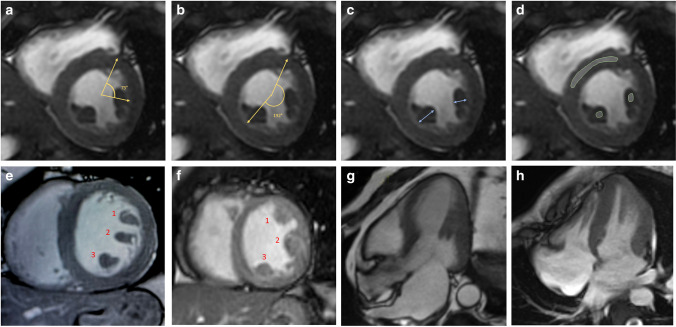
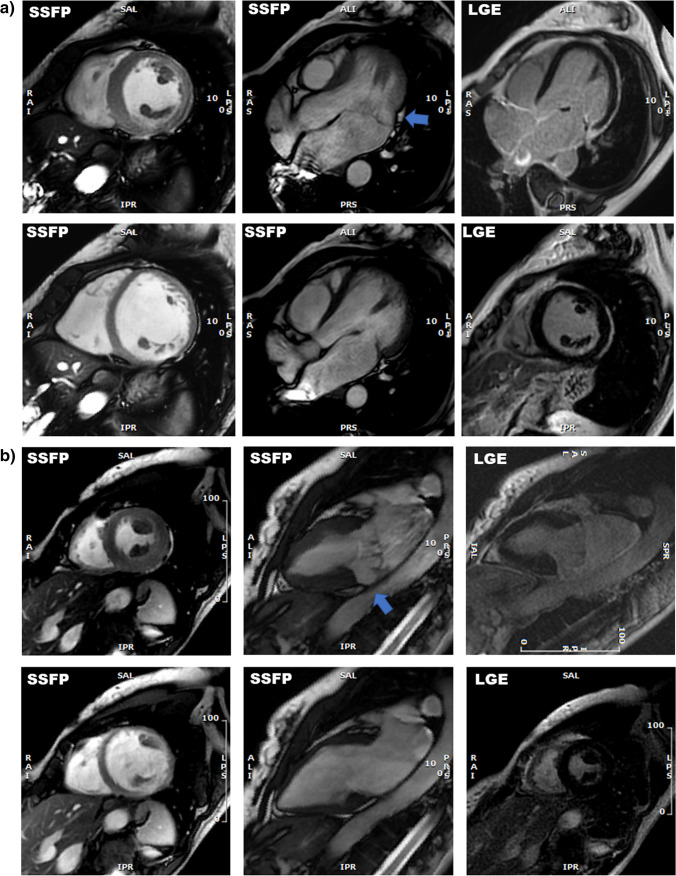
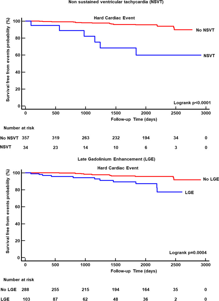
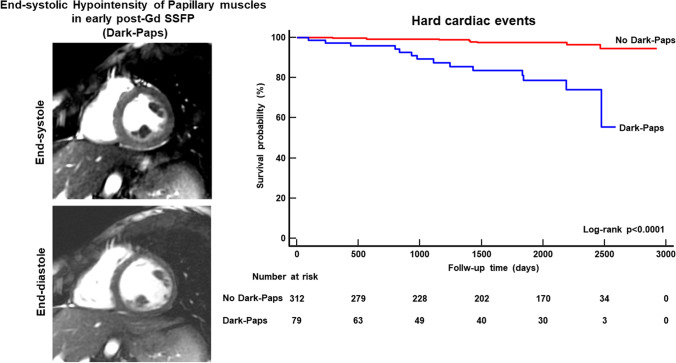
Methods: A total of 391 patients with > 500/24 h premature ventricular complexes and/or with non-sustained ventricular tachycardia (NSVT), preserved LVEF, and no cardiac disease were enrolled. Different features of LV papillary muscles were considered: supernumerary muscles, papillary thickness, the attachment, late gadolinium enhancement (LGE). Dark-Paps was defined as end-systolic signal hypointensity of both papillary muscles in early post-contrast cine CMR images. Mitral valve prolapse, mitral annular disjunction (MAD), and myocardial LGE were considered.
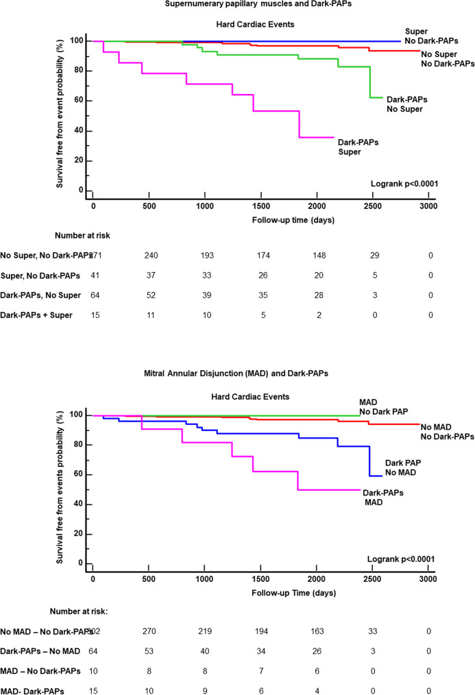
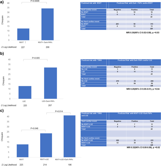
Results: Dark-Paps was found in 79 (20%) patients and was more frequent in females. It was associated with higher prevalence of mitral valve prolapse and MAD. During a median follow-up of 2534 days, 22 hard cardiac events occurred. At Kaplan-Meier curve analysis, patients with Dark-Paps were at higher risk of events than those without (p < 0.0001). Dark-Paps was significantly associated with hard cardiac events in all the multivariate models. Dark-Paps improved prognostic estimation when added to NSVT (p = 0.0006), to LGE (p = 0.005) and to a model including NSVT+LGE (p = 0.014). Dark-Paps allowed a significant net reclassification when added to NSVT (NRI 0.30, p = 0.03), to LGE (NRI 0.25, p = 0.04), and to NSVT + LGE (NRI 0.32, p = 0.02).
Conclusions: In LV papillary muscles, Dark-Paps is a novel prognostic marker in patients with ventricular arrhythmias and preserved ejection fraction.
Key points: • Papillary muscle abnormalities are seen in patients with ventricular arrhythmias and preserved left ventricular ejection fraction. • Early post-contrast hypointensity of papillary muscles in end-systolic cine images (Dark-Paps) is a novel prognostic marker in patients with ventricular arrhythmias and preserved ejection fraction. • Dark-Paps had an additive prognostic role over late gadolinium enhancement and non-sustained ventricular tachycardia.
Keywords: Cardiac magnetic resonance; Papillary muscles; Prognosis; Sudden cardiac death.
© 2023. The Author(s).
Conflict of interest statement
The authors of this manuscript declare no relationships with any companies whose products or services may be related to the subject matter of the article.
Figures
References
-
- Harrigan CJ, Appelbaum E, Maron BJ et al (2008) Significance of papillary muscle abnormalities identified by cardiovascular magnetic resonance in hypertrophic cardiomyopathy. Am J Cardiol 101:668–673 - PubMed
-
- Maron MS, Olivotto I, Harrigan C et al (2011) Mitral valve abnormalities identified by cardiovascular magnetic resonance represent a primary phenotypic expression of hypertrophic cardiomyopathy. Circulation 124:40–47 - PubMed
MeSH terms
Substances
LinkOut - more resources
Full Text Sources
Medical
