

Molecular characterization of eliminated chromosomes in Hessian fly (Mayetiola destructor (Say))
- PMID: 36692656
- PMCID: PMC9873768
- DOI: 10.1007/s10577-023-09718-8
Molecular characterization of eliminated chromosomes in Hessian fly (Mayetiola destructor (Say))
Abstract
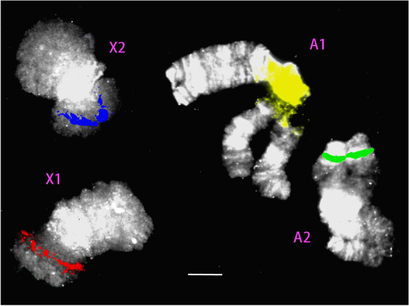
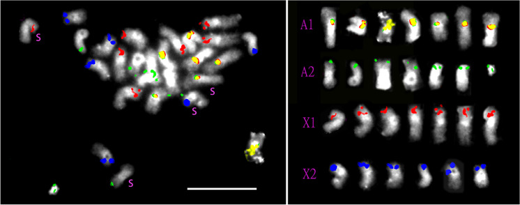
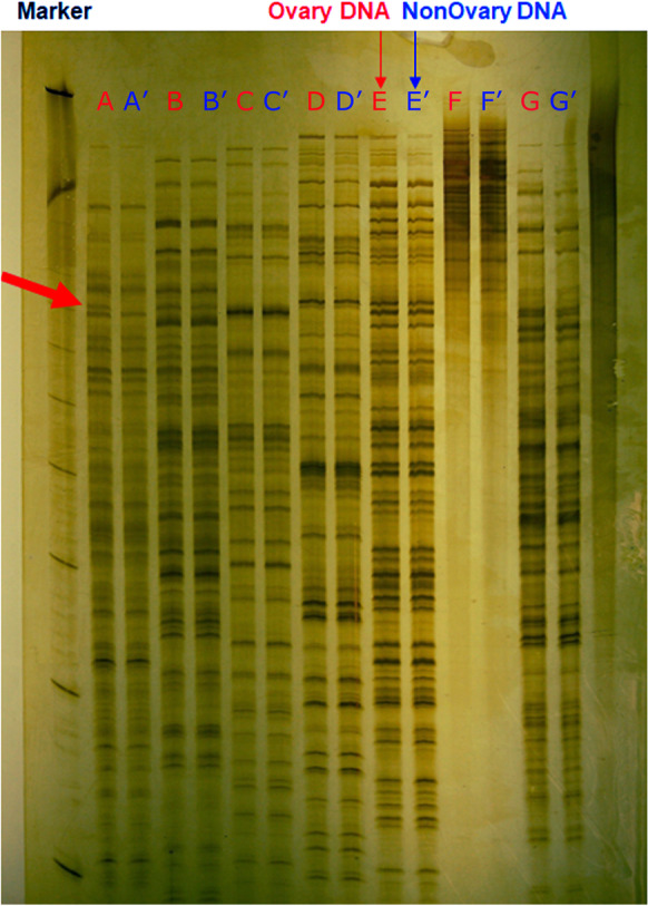
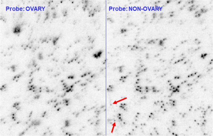
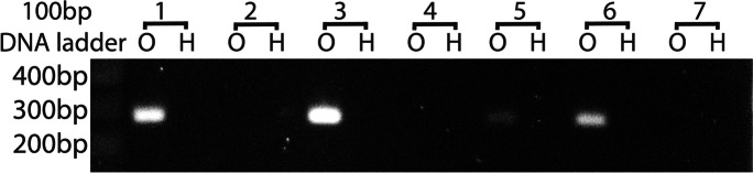
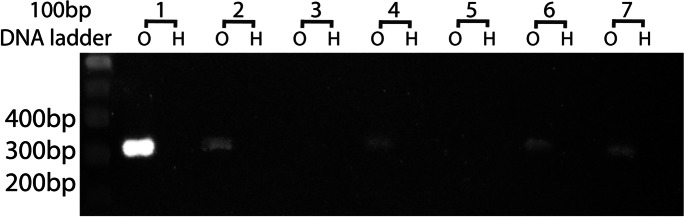
Like other cecidomyiid Diptera, Hessian fly has stable S chromosomes and dispensable E chromosomes that are retained only in the germ line. Amplified fragment length polymorphisms (AFLP), suppressive subtractive hybridization (SSH), fluorescent in-situ hybridization (FISH), and sequencing were used to investigate similarities and differences between S and E chromosomes. More than 99.9% of AFLP bands were identical between separated ovary and somatic tissue, but one band was unique to ovary and resembled Worf, a non-LTR retrotransposon. Arrayed clones, derived by SSH of somatic from ovarian DNA, showed no clones that were unique to ovary. FISH with BAC clones revealed a diagnostic banding pattern of BAC positions on both autosomes and both sex chromosomes, and each E chromosome shared a pattern with one of the S chromosomes. Sequencing analysis showed that E chromosomes are nearly identical to S chromosomes, since no sequence could be confirmed to belong only to E chromosomes. There were a few questionably E-specific sequences that are candidates for further investigation. Thus, the E chromosomes appear to be derived from S chromosomes by the acquisition or conversion of sequences that produce the negatively heteropycnotic region around the centromere.
Keywords: DNA sequencing; Hessian fly; chromosome elimination; germline; supernumerary chromosome.
© 2023. This is a U.S. Government work and not under copyright protection in the US; foreign copyright protection may apply.
Conflict of interest statement
The authors declare no conflicts of interest regarding the publication of this paper. Mention of a commercial or proprietary product does not constitute an endorsement by the US Federal Government.
Figures

Similar articles
-
A BAC-based physical map of the Hessian fly genome anchored to polytene chromosomes.BMC Genomics. 2009 Jul 2;10:293. doi: 10.1186/1471-2164-10-293. BMC Genomics. 2009. PMID: 19573234 Free PMC article.
-
Localization and characterization of 170 BAC-derived clones and mapping of 94 microsatellites in the Hessian fly.J Hered. 2009 Nov-Dec;100(6):790-7. doi: 10.1093/jhered/esp045. Epub 2009 Jul 10. J Hered. 2009. PMID: 19592640
-
Assessment of structural variation and molecular mapping of insertion sites of Desmar-like elements in the Hessian fly genome.Insect Mol Biol. 2010 Dec;19(6):707-15. doi: 10.1111/j.1365-2583.2010.01028.x. Insect Mol Biol. 2010. PMID: 20636348
-
A neo-sex chromosome that drives postzygotic sex determination in the hessian fly (Mayetiola destructor).Genetics. 2010 Mar;184(3):769-77. doi: 10.1534/genetics.109.108589. Epub 2009 Dec 21. Genetics. 2010. PMID: 20026681 Free PMC article.
-
Genetic and cytogenetic analysis of the olive fruit fly Bactrocera oleae (Diptera: Tephritidae).Genetica. 2002 Sep;116(1):45-57. doi: 10.1023/a:1020907624816. Genetica. 2002. PMID: 12484525 Review.
Cited by
-
Functions and mechanisms of eukaryotic RNA-guided programmed DNA elimination.Biochem Soc Trans. 2025 Apr 29;53(2):473-85. doi: 10.1042/BST20253006. Biochem Soc Trans. 2025. PMID: 40305257 Free PMC article. Review.
References
-
- Bankevich A, Nurk S, Antipov D, Gurevich AA, Dvokin M, Kulikov AS, Lesin VM, Nikolenko SI, Pham S, Prjibelski AD, Pyshkin AV, Sirotkin AV, Vyahhi N, Tesler G, Alekseyev MA, Pevzner PA. SPAdes: a new genome assembly algorithm and its applications to single-cell sequencing. J Comp Biol. 2012;19(5):455–477. doi: 10.1089/cmb.2012.0021. - DOI - PMC - PubMed
-
- Bantock CR. Experiments on chromosome elimination in the gall midge, Mayetiola destructor. J Embryol Exp Morpholog. 1970;24:257–286. - PubMed
Publication types
MeSH terms
Substances
LinkOut - more resources
Full Text Sources
