Knee positioning systems for X-ray environment: a literature review
- PMID: 36692683
- PMCID: PMC10030541
- DOI: 10.1007/s13246-023-01221-y
Knee positioning systems for X-ray environment: a literature review
Abstract
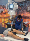
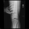
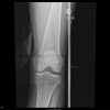
The knee is one of the most stressed joints of the human body, being susceptible to ligament injuries and degenerative diseases. Due to the rising incidence of knee pathologies, the number of knee X-rays acquired is also increasing. Such X-rays are obtained for the diagnosis of knee injuries, the evaluation of the knee before and after surgery, and the monitoring of the knee joint's stability. These types of diagnosis and monitoring of the knee usually involve radiography under physical stress. This widely used medical tool provides a more objective analysis of the measurement of the knee laxity than a physical examination does, involving knee stress tests, such as valgus, varus, and Lachman. Despite being an improvement to physical examination regarding the physician's bias, stress radiography is still performed manually in a lot of healthcare facilities. To avoid exposing the physician to radiation and to decrease the number of X-ray images rejected due to inadequate positioning of the patient or the presence of artefacts, positioning systems for stress radiography of the knee have been developed. This review analyses knee positioning systems for X-ray environment, concluding that they have improved the objectivity and reproducibility during stress radiographs, but have failed to either be radiolucent or versatile with a simple ergonomic set-up.
Keywords: Knee joint; Lachman test; Positioning system; Stress radiography; Valgus stress; Varus stress.
© 2023. The Author(s).
Conflict of interest statement
All authors certify that they have no affiliations with or involvement in any organization or entity with any financial interest or non-financial interest in the subject matter or materials discussed in this manuscript.
Figures


Similar articles
-
Good clinical and radiological results of total knee arthroplasty using varus valgus constrained or rotating hinge implants in ligamentous laxity.Knee Surg Sports Traumatol Arthrosc. 2019 May;27(5):1665-1670. doi: 10.1007/s00167-018-5307-6. Epub 2018 Nov 20. Knee Surg Sports Traumatol Arthrosc. 2019. PMID: 30456570
-
Reliability of stress radiography in the assessment of coronal laxity following total knee arthroplasty.Knee. 2020 Jan;27(1):221-228. doi: 10.1016/j.knee.2019.09.013. Epub 2019 Dec 23. Knee. 2020. PMID: 31875838
-
Stress Radiographs for Ligamentous Knee Injuries.Arthroscopy. 2021 Jan;37(1):15-16. doi: 10.1016/j.arthro.2020.11.001. Arthroscopy. 2021. PMID: 33384079
-
Is knee radiography useful for studying the efficacy of a disease-modifying osteoarthritis drug in humans?Rheum Dis Clin North Am. 2003 Nov;29(4):819-30. doi: 10.1016/s0889-857x(03)00055-3. Rheum Dis Clin North Am. 2003. PMID: 14603585 Review.
-
Imaging Review of the Posterior Cruciate Ligament.J Knee Surg. 2021 Apr;34(5):493-498. doi: 10.1055/s-0040-1722629. Epub 2021 Feb 22. J Knee Surg. 2021. PMID: 33618404 Review.
Cited by
-
Knee Osteoarthritis Diagnosis: Future and Perspectives.Biomedicines. 2025 Jul 4;13(7):1644. doi: 10.3390/biomedicines13071644. Biomedicines. 2025. PMID: 40722715 Free PMC article. Review.
-
Ultrasound-based bone tracking using cross-correlation enables dynamic measurements of knee kinematics during clinical assessments.J Exp Orthop. 2024 Jun 6;11(3):e12050. doi: 10.1002/jeo2.12050. eCollection 2024 Jul. J Exp Orthop. 2024. PMID: 38846378 Free PMC article.
-
Is There a Role of Photoacoustic Imaging in Sports Medicine: Evidence Today.Orthop Surg. 2025 Jun;17(6):1589-1603. doi: 10.1111/os.70031. Epub 2025 Mar 25. Orthop Surg. 2025. PMID: 40132986 Free PMC article. Review.
-
Effects of photobiomodulation combined with rehabilitation exercise on pain, physical function, and radiographic changes in mild to moderate knee osteoarthritis: A randomized controlled trial protocol.PLoS One. 2025 Jan 21;20(1):e0314869. doi: 10.1371/journal.pone.0314869. eCollection 2025. PLoS One. 2025. PMID: 39836628 Free PMC article.
-
Hydrogel composite scaffold repairs knee cartilage defects: a systematic review.RSC Adv. 2025 Apr 8;15(13):10337-10364. doi: 10.1039/d5ra01031d. eCollection 2025 Mar 28. RSC Adv. 2025. PMID: 40200956 Free PMC article. Review.
References
-
- American Cancer Society (N.D.) Do X-rays and gamma rays cause any other health problems? American Cancer Society. https://www.cancer.org/healthy/cancer-causes/radiation-exposure/x-rays-g.... Accessed 29 Nov 2022
-
- Anterior cruciate ligament injury. https://somepomed.org/articulos/contents/mobipreview.htm?31/59/32689. Accessed 28 Nov 2022
-
- Beaumont Health (N.D.) Sports injuries | anterior cruciate ligament (ACL) tears. Beaumont Health. https://www.beaumont.org/conditions/acl-tears. Accessed 28 Nov 2022
Publication types
MeSH terms
LinkOut - more resources
Full Text Sources
