. 2023 May;60(5):2602-2618.
doi: 10.1007/s12035-023-03219-9. Epub 2023 Jan 24.

A Homozygous PPP1R21 Splice Variant Associated with Severe Developmental Delay, Absence of Speech, and Muscle Weakness Leads to Activated Proteasome Function

Affiliations

A Homozygous PPP1R21 Splice Variant Associated with Severe Developmental Delay, Absence of Speech, and Muscle Weakness Leads to Activated Proteasome Function

Andreas Hentschel et al. Mol Neurobiol. 2023 May.

Erratum in

Abstract

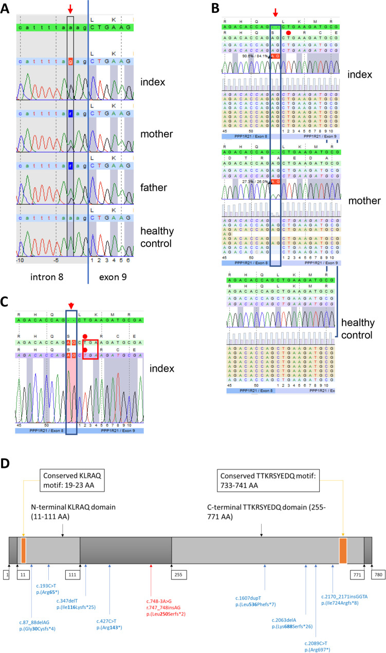
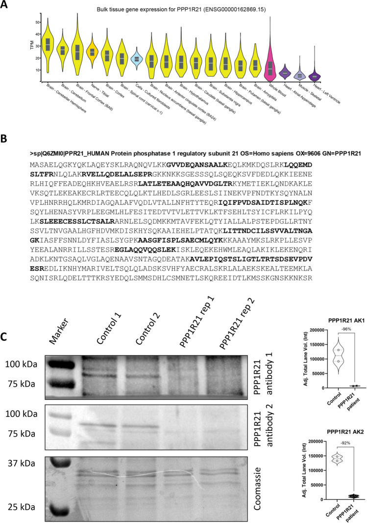
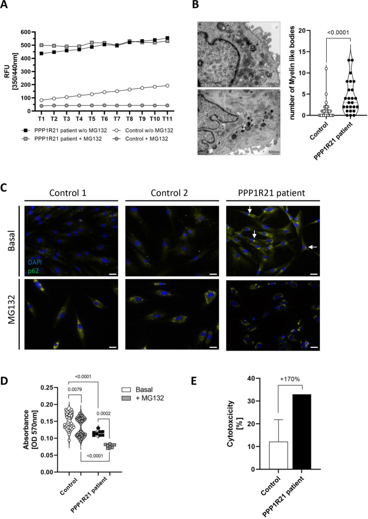
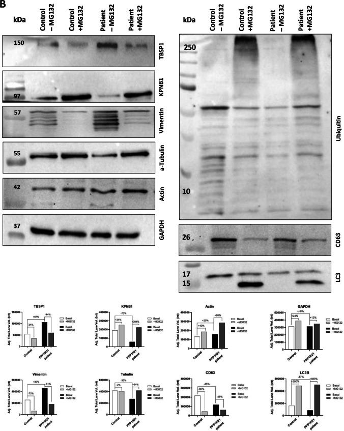
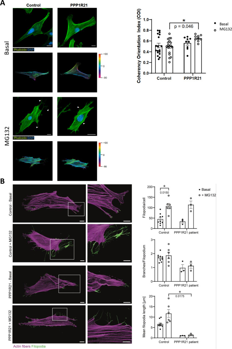
PPP1R21 acts as a co-factor for protein phosphatase 1 (PP1), an important serine/threonine phosphatase known to be essential for cell division, control of glycogen metabolism, protein synthesis, and muscle contractility. Bi-allelic pathogenic variants in PPP1R21 were linked to a neurodevelopmental disorder with hypotonia, facial dysmorphism, and brain abnormalities (NEDHFBA) with pediatric onset. Functional studies unraveled impaired vesicular transport as being part of PPP1R21-related pathomechanism. To decipher further the pathophysiological processes leading to the clinical manifestation of NEDHFBA, we investigated the proteomic signature of fibroblasts derived from the first NEDHFBA patient harboring a splice-site mutation in PPP1R21 and presenting with a milder phenotype. Proteomic findings and further functional studies demonstrate a profound activation of the ubiquitin-proteasome system with presence of protein aggregates and impact on cellular fitness and moreover suggest a cross-link between activation of the proteolytic system and cytoskeletal architecture (including filopodia) as exemplified on paradigmatic proteins including actin, thus extending the pathophysiological spectrum of the disease. In addition, the proteomic signature of PPP1R21-mutant fibroblasts displayed a dysregulation of a variety of proteins of neurological relevance. This includes increase proteins which might act toward antagonization of cellular stress burden in terms of pro-survival, a molecular finding which might accord with the presentation of a milder phenotype of our NEDHFBA patient.

Keywords: Fibroblast cytoskeleton; Fibroblast electron microscopy; Fibroblast filopodia; Fibroblast proteomics; NEDHFBA; PPP1R21; Proteasome.


Conflict of interest statement

Seven of the authors of this publication are members of the European Reference Network for Neuromuscular Diseases-Project ID N° 870177. Three authors of this study are part of the Center for Translational Neuro- and Behavioral Sciences (C-NTBS). The other authors declare no competing interests.

Figures

Fig. 1
Fig. 1
Molecular genetic findings. A Sanger sequencing of genomic DNA extracted from peripheral blood confirmed the splice site variant at position − 3 of intron 8 of the PPP1R21 gene (c.748-3A > G) presenting as homozygous in the index patient and heterozygous in both parents, respectively. A wild-type sequence of a healthy control individual is shown in the lower panel. B Next generation sequencing of PPP1R21 cDNA obtained from RNA isolated from peripheral blood samples of the index patient, the mother, and a healthy control; utilization of an alternative acceptor site leads to an insertion of two nucleotides (r.747_748insAG) between exons 8 and 9 of the resulting mRNA is seen in the index patient and his mother. Due to this insertion, the frameshift results in a premature stop codon at the beginning of exon 9 (p.Leu250Serfs*2). C Sanger sequencing of cDNA obtained from RNA isolated from cultured fibroblasts of the index patient confirms the insertion of two nucleotides between the exons 8 and 9 resulting in a frame shift and a premature stop codon at the beginning of exon 9 (p.Leu250Serfs*2). D Illustration of PPP1R21 structure including labeling of mutation site(s) on both, the nucleic acid and the amino acid (AA) level. Mutations reported previously (see references [4, 7, 8]) are labeled in blue whereas the pathogenic variant identified in our patient is labeled in red. Numbers in boxes refer to amino acid positions
Fig. 2
Fig. 2
PPP1R21 expression in human fibroblasts. A GTEx-based in silico analysis of PPP1R21 revealed expression in nervous tissues (yellow), cultured fibroblasts (blue), blood (pink), skeletal (light purple), and cardiac muscle (dark purple). B Sequence coverage of PPP1R21 based on unique peptides catalog in our spectral library. C Immunoblot studies (including quantification) utilizing two anti-PPP1R21 antibodies on whole protein extracts show loss of PPP1R21 protein abundance in patient-derived fibroblasts compared to controls. Coomassie staining is shown to show equal protein loading
Fig. 3
Fig. 3
Proteomic profiling on PPP1R21 patient fibroblasts. A Volcano plot highlighting statistically significant increased proteins (green dots) as well as decreased proteins (red dots). B Box plots of selected increased proteins including ATXN (ataxin-10), SNTB1 (Beta-1-synthrophin), CDR2L (cerebellar degeneration-related protein 2-like), SLC1A3 (excitatory amino acid transporter 1), FAM126A (hyccin), PPP1R9B (neurabin-2), NCS1 (neuronal calcium sensor 1), SACS (sacsin), and SVIL (supervillin) to visualize affection of neuronal and neuromuscular relevant proteins based on loss of functional PPP1R21 in cultured human fibroblasts. C Proteomaps-based analysis to decipher the impact on dysregulated proteins (left panel) on diverse biological and cellular processes (right panel). This analysis was performed for upregulated (upper panel) and downregulated (lower panel) proteins, respectively
Fig. 4
Fig. 4
Cellular studies on the functional and microscopic level. A Investigation of proteasomal activity shows a five-fold increase of proteasomal function in PPP1R21 patient-derived fibroblasts compared to pooled controls under basal conditions. After treatment with MG132 (10 µM for 16 h), control cells do not show proteasomal activity whereas there is no effect of treatment in fibroblasts lacking PPP1R21. B Ultrastructural analysis of cultured fibroblasts; only single myelin figures are present in the cytoplasm of control fibroblast as exemplified in the patient-derived (upper panel) and control cells (lower panel). In comparison to two controls analyzed, in PPP1R21-mutant fibroblast number of myelin-like bodies is increased as shown by counting respective numbers presented in the box plots (right panel). C Immunofluorescence studies on PPP1R21 patient derived and control fibroblasts showed increase of p62 in the perinuclear region in patient derived but not in control cells under basal conditions (white arrows). After treatment of cells with MG132, increase of p62-immunoreactivity is detectable at similar level in fibroblasts derived from controls and the PPP1R21 patient. Scale bars: 20 µm. D Studies of cellular fitness by MTT-assay revealed mild reduction of cellular viability mirrored by the metabolic state and membrane permeability in patient-derived cells compared to matched controls. The impact of inhibition of proteasomal activity on cellular viability by MG132 treatment was more pronounced in patient-derived cells compared to controls. E A more pronounced increase of cytotoxicity after MG132-treatment is present in patient-derived cells compared to control fibroblasts
Fig. 5
Fig. 5
Transcript and immunoblot studies. A Quantitative PCR studies of selected transcripts for UPR-modulators (ATF4, ATF6, BiP, CALR, CALU, CANX, EDEM1, EIF2A, GRP94, GRP170, IRE1, PDIA3, PERK, and XBP1) and proteolysis (BAG3, BECN1, HSPA8, HSPB8, LAMP1, LC3, LMP7, PSME1, and PSME2) revealed a clear effect of MG132 on expression levels of UPR-related genes in control fibroblast. Furthermore, PPP1R21 patient and control fibroblasts present with comparable expression level under basal control expression, with exception of BiP and GRP170. Treatment with MG132 decreased the expression of both UPR-modulators, while other markers showed elevated expression. PERK, XBP1, and GRP94 only show increase upon MG132 treatment in control but not patient-derived fibroblasts. Among autophagy modulating factors, BECLN, LC3, and HSPB8 as well as the transcripts LMP7, PSME1, and PSME2 encoding for proteasomal proteins were increased upon MG132 treatment in both, patient derived and control fibroblasts. None of the autophagy modulating factors or proteasomal protein show an increase under basal conditions in patient-derived fibroblasts. B Immunoblot studies (including quantification) focusing on proteins belonging to cytoskeleton (alpha-tubulin, vimentin, and actin), KPNB1 as being identified to be dysregulated by proteomic profiling, and such involved in protein clearance (ubiquitin, CD63, and LC3) confirmed affection of cytoskeleton and protein degradation in fibroblasts lacking expression of PPP1R21 and moreover show alterations of these processes in cells treated with MG132 by the detection of increase of vimentin, actin, and ubiquitin and reduced conversion from LC3-I to LC3-II
Fig. 5
Fig. 5
Transcript and immunoblot studies. A Quantitative PCR studies of selected transcripts for UPR-modulators (ATF4, ATF6, BiP, CALR, CALU, CANX, EDEM1, EIF2A, GRP94, GRP170, IRE1, PDIA3, PERK, and XBP1) and proteolysis (BAG3, BECN1, HSPA8, HSPB8, LAMP1, LC3, LMP7, PSME1, and PSME2) revealed a clear effect of MG132 on expression levels of UPR-related genes in control fibroblast. Furthermore, PPP1R21 patient and control fibroblasts present with comparable expression level under basal control expression, with exception of BiP and GRP170. Treatment with MG132 decreased the expression of both UPR-modulators, while other markers showed elevated expression. PERK, XBP1, and GRP94 only show increase upon MG132 treatment in control but not patient-derived fibroblasts. Among autophagy modulating factors, BECLN, LC3, and HSPB8 as well as the transcripts LMP7, PSME1, and PSME2 encoding for proteasomal proteins were increased upon MG132 treatment in both, patient derived and control fibroblasts. None of the autophagy modulating factors or proteasomal protein show an increase under basal conditions in patient-derived fibroblasts. B Immunoblot studies (including quantification) focusing on proteins belonging to cytoskeleton (alpha-tubulin, vimentin, and actin), KPNB1 as being identified to be dysregulated by proteomic profiling, and such involved in protein clearance (ubiquitin, CD63, and LC3) confirmed affection of cytoskeleton and protein degradation in fibroblasts lacking expression of PPP1R21 and moreover show alterations of these processes in cells treated with MG132 by the detection of increase of vimentin, actin, and ubiquitin and reduced conversion from LC3-I to LC3-II
Fig. 6
Fig. 6
Studies of actin cytoskeleton. A Actin filaments (Phalloidin, green) and nuclei (DAPI, blue) of control and PPP1R21 patient-derived fibroblasts are visualized via confocal laser scanning microscopy. MG132 treated fibroblasts of control patients exhibit longer and more branched filopodia (white arrows) than PPP1R21 patient fibroblasts. Scale bars: 20 µm. Circular color map coding displays the local actin filament orientation of the basal and MG132 treated fibroblasts derived from controls and the PPP1R21 patient, respectively. The color gradient indicates the spatial orientation of the actin fibers from − 90° to + 90°. MG132 treatment induced a significant increase in the cytoskeletal coherency of PPP1R21 patient fibroblasts compared to stimulated controls. B Microscopic studies of fibroblast filopodia (illustrated in green) revealed an effect of MG132 exposure in terms of an increase of filopodia formation per cell in both, control- and patient-derived cells. Branches and length per filopodia are (slightly) reduced in patient-derived cells and. Mean filopodia length increased upon MG132 treatment only in control fibroblasts. Scale bars in overview images = 20 µm, scale bars in magnification = 10 µm

Similar articles

Cited by

References

    1. Peel N, Iyer J, Naik A, et al. Protein phosphatase 1 down regulates ZYG-1 levels to limit centriole duplication. PLoS Genet. 2017;13:e1006543. doi: 10.1371/journal.pgen.1006543. - DOI - PMC - PubMed
    1. Munro S, Cuthbertson DJR, Cunningham J, et al. Human skeletal muscle expresses a glycogen-targeting subunit of PP1 that is identical to the insulin-sensitive glycogen-targeting subunit G(L) of liver. Diabetes. 2002;51:591–598. doi: 10.2337/diabetes.51.3.591. - DOI - PubMed
    1. Strack S, Kini S, Ebner FF, et al. Differential cellular and subcellular localization of protein phosphatase 1 isoforms in brain. J Comp Neurol. 1999;413:373–384. doi: 10.1002/(SICI)1096-9861(19991025)413:3<373::AID-CNE2>3.0.CO;2-Z. - DOI - PubMed
    1. Rehman AU, Najafi M, Kambouris M, et al. Biallelic loss of function variants in PPP1R21 cause a neurodevelopmental syndrome with impaired endocytic function. Hum Mutat. 2019;40:267–280. doi: 10.1002/humu.23694. - DOI - PMC - PubMed
    1. Anazi S, Maddirevula S, Salpietro V, et al. Expanding the genetic heterogeneity of intellectual disability. Hum Genet. 2017;136:1419–1429. doi: 10.1007/s00439-017-1843-2. - DOI - PubMed

LinkOut - more resources