

Clonorchis sinensis aggravates biliary fibrosis through promoting IL-6 production via toll-like receptor 2-mediated AKT and p38 signal pathways
- PMID: 36693049
- PMCID: PMC9873171
- DOI: 10.1371/journal.pntd.0011062
Clonorchis sinensis aggravates biliary fibrosis through promoting IL-6 production via toll-like receptor 2-mediated AKT and p38 signal pathways
Abstract
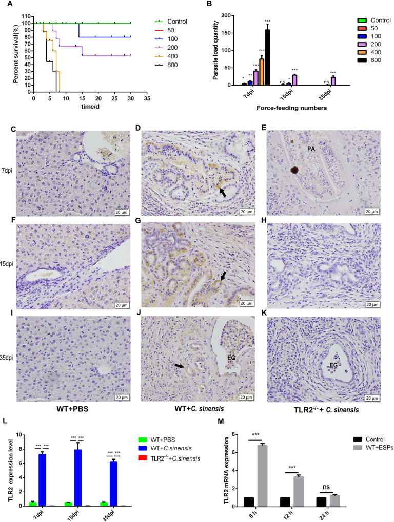
Clonorchis sinensis is an important food-borne zoonotic parasite which has been linked to biliary fibrosis and cholangiocarcinoma. However, the details of the pathogenesis of C. sinensis were unclear. To explore the role and regulatory mechanism of toll-like receptor 2 (TLR2) in C. sinensis-induced biliary fibrosis, we established the C. sinensis-infected C57BL/6 mouse model with TLR2-/- and wild type (WT) mice. The mortality rate, liver lesions, TLR2 and TGF-β1 expression, phosphorylation of Smad2/3, AKT, p38, ERK and p65, and cytokine productions were analyzed. Furthermore, similar parameters were examined in mouse biliary epithelial cells (BECs) co-cultured with C. sinensis excretory/secretory proteins (ESPs). The results showed that TLR2 expression was enhanced significantly in C. sinensis-infected WT mice and mouse BECs. C. sinensis-infected TLR2-/- mice exhibited an increased weight and a decreased mortality rate; significantly alleviated liver lesions and biliary fibrosis, reduced numbers of myofibroblasts; decreased expression of TGF-β1 and phosphorylation level of AKT, p38 and Smad2/3; significantly decreased production of IL-6, TNF-α and IL-4, while increased production of IFN-γ compared with C. sinensis-infected WT mice. Furthermore, C. sinensis ESPs could activate TLR2-mediated AKT and p38 pathways to increase the production of IL-6 in mouse BECs. In conclusion, these data indicate that C. sinensis infection activated TGF-β1-Smad2/3 through TLR2-mediated AKT and p38 pathways to promote IL-6 production, which resulted in myofibroblast activation and aggravating biliary fibrosis in mice.
Copyright: © 2023 Wang et al. This is an open access article distributed under the terms of the Creative Commons Attribution License, which permits unrestricted use, distribution, and reproduction in any medium, provided the original author and source are credited.
Conflict of interest statement
The authors have declared that no competing interests exist.
Figures


Similar articles
-
TLR3 activation by Clonorchis sinensis infection alleviates the fluke-induced liver fibrosis.PLoS Negl Trop Dis. 2023 May 11;17(5):e0011325. doi: 10.1371/journal.pntd.0011325. eCollection 2023 May. PLoS Negl Trop Dis. 2023. PMID: 37167198 Free PMC article.
-
The crosstalk between cholangiocytes and hepatic stellate cells promotes the progression of epithelial-mesenchymal transition and periductal fibrosis during Clonorchis sinensis infection.Parasit Vectors. 2024 Mar 22;17(1):151. doi: 10.1186/s13071-024-06236-2. Parasit Vectors. 2024. PMID: 38519993 Free PMC article.
-
TLR2 signal influences the iNOS/NO responses and worm development in C57BL/6J mice infected with Clonorchis sinensis.Parasit Vectors. 2017 Aug 7;10(1):379. doi: 10.1186/s13071-017-2318-y. Parasit Vectors. 2017. PMID: 28784165 Free PMC article.
-
Clonorchis sinensis and clonorchiasis.Acta Trop. 2020 Mar;203:105309. doi: 10.1016/j.actatropica.2019.105309. Epub 2019 Dec 17. Acta Trop. 2020. PMID: 31862466 Review.
-
Vibrio harveyi infections induce production of proinflammatory cytokines in murine peritoneal macrophages via activation of p38 MAPK and NF-κB pathways, but reversed by PI3K/AKT pathways.Dev Comp Immunol. 2022 Feb;127:104292. doi: 10.1016/j.dci.2021.104292. Epub 2021 Oct 14. Dev Comp Immunol. 2022. PMID: 34656643 Review.
Cited by
-
Helminth-derived molecules: Pathogenic and pharmacopeial roles.J Biomed Res. 2024 Sep 24;38(6):547-568. doi: 10.7555/JBR.38.20240177. J Biomed Res. 2024. PMID: 39314046 Free PMC article.
-
TLR3 activation by Clonorchis sinensis infection alleviates the fluke-induced liver fibrosis.PLoS Negl Trop Dis. 2023 May 11;17(5):e0011325. doi: 10.1371/journal.pntd.0011325. eCollection 2023 May. PLoS Negl Trop Dis. 2023. PMID: 37167198 Free PMC article.
-
Biliary fibrosis is an important but neglected pathological feature in hepatobiliary disorders: from molecular mechanisms to clinical implications.Med Rev (2021). 2024 Jul 1;4(4):326-365. doi: 10.1515/mr-2024-0029. eCollection 2024 Aug. Med Rev (2021). 2024. PMID: 39135601 Free PMC article. Review.
-
Hepatic ferroptosis induced by Clonorchis sinensis exacerbates liver fibrosis.PLoS Negl Trop Dis. 2025 Jun 2;19(6):e0013164. doi: 10.1371/journal.pntd.0013164. eCollection 2025 Jun. PLoS Negl Trop Dis. 2025. PMID: 40455823 Free PMC article.
-
Prevalence and species identification of trematode metacercariae in Qiqihar, Northeast China.Front Microbiol. 2024 Sep 10;15:1464988. doi: 10.3389/fmicb.2024.1464988. eCollection 2024. Front Microbiol. 2024. PMID: 39318428 Free PMC article.
References
-
- WHO. Working to overcome the global impact of neglected tropical diseases: First WHO report on neglected tropical diseases. Geneva World Heal Organ 2010. 148: 148–162. https://www.who.int/publications-detail-redirect/9789241564090
-
- Hong ST, Huh S, Kho WG, Yu JR, Chai JY, Kim EC, et al.. Changes in histopathological and serological findings of the liver after treatment in rabbit clonorchiasis. Seoul J Med. 1990; 117–127. https://s-space.snu.ac.kr/bitstream/10371/7310/1/SeoulJMed31.117-127.pdf
Publication types
MeSH terms
Substances
LinkOut - more resources
Full Text Sources
Medical
Miscellaneous
