. 2023 Jan 31;120(5):e2214684120.
doi: 10.1073/pnas.2214684120. Epub 2023 Jan 24.

METTL3 is essential for normal progesterone signaling during embryo implantation via m6A-mediated translation control of progesterone receptor

Affiliations

METTL3 is essential for normal progesterone signaling during embryo implantation via m6A-mediated translation control of progesterone receptor

Zhan-Hong Zheng et al. Proc Natl Acad Sci U S A. .

Abstract

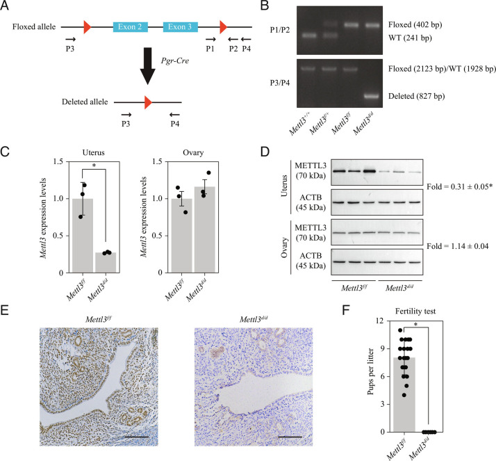
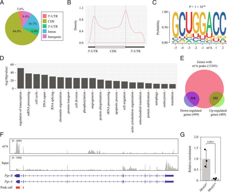
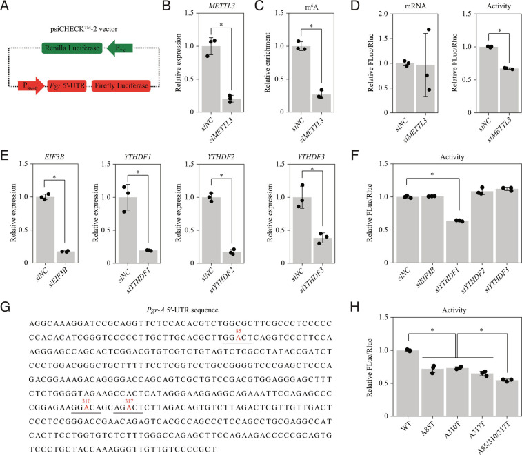
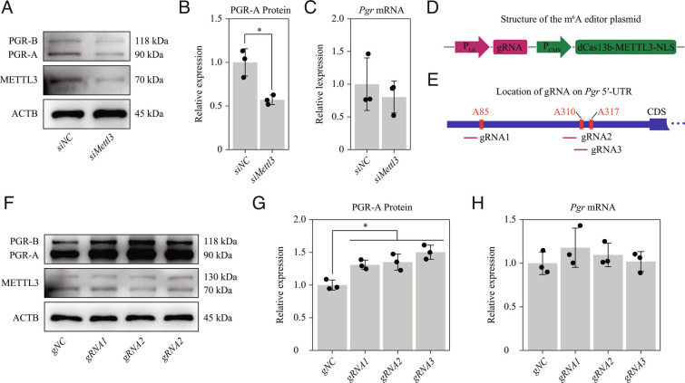
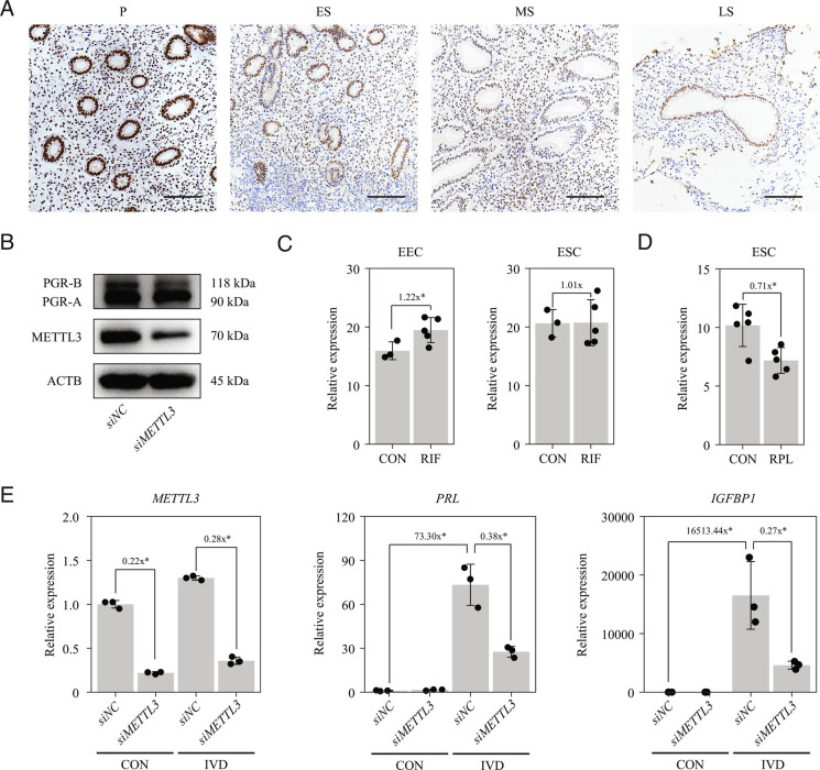
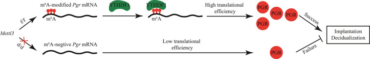
Embryo implantation, a crucial step in human reproduction, is tightly controlled by estrogen and progesterone (P4) via estrogen receptor alpha and progesterone receptor (PGR), respectively. Here, we report that N6-methyladenosine (m6A), the most abundant mRNA modification in eukaryotes, plays an essential role in embryo implantation through the maintenance of P4 signaling. Conditional deletion of methyltransferase-like 3 (Mettl3), encoding the m6A writer METTL3, in the female reproductive tract using a Cre mouse line with Pgr promoter (Pgr-Cre) resulted in complete implantation failure due to pre-implantation embryo loss and defective uterine receptivity. Moreover, the uterus of Mettl3 null mice failed to respond to artificial decidualization. We further found that Mettl3 deletion was accompanied by a marked decrease in PGR protein expression. Mechanistically, we found that Pgr mRNA is a direct target for METTL3-mediated m6A modification. A luciferase assay revealed that the m6A modification in the 5' untranslated region (5'-UTR) of Pgr mRNA enhances PGR protein translation efficiency in a YTHDF1-dependent manner. Finally, we demonstrated that METTL3 is required for human endometrial stromal cell decidualization in vitro and that the METTL3-PGR axis is conserved between mice and humans. In summary, this study provides evidence that METTL3 is essential for normal P4 signaling during embryo implantation via m6A-mediated translation control of Pgr mRNA.

Keywords: METTL3; embryo implantation; m6A; progesterone receptor.


Conflict of interest statement

The authors declare no competing interest.

Figures

Fig. 1.
Fig. 1.
Pgr-Cre-mediated deletion of Mettl3 leads to complete infertility. (A) Diagram showing the strategy used for conditional Mettl3 deletion. Mettl3 floxed (Mettl3f/f) mice have loxP sites flanking exons 2 and 3. Conditional Mettl3 deletion (Mettl3d/d) mice were generated by crossing Mettl3f/f mice with mice of the Pgr-Cre driver line. (B) Genotyping analysis showing the Mettl3 knockout efficiency at the DNA level in the uterus on gestational day 4 (GD4). (C) Quantitative RT-PCR analysis of Mettl3 mRNA levels in the uterus and the ovary of Mettl3f/f and Mettl3d/d mice on GD4. Data are presented as mean ± SD. *P < 0.05. (D) Western blot analysis of METTL3 protein levels in the uterus and the ovary on GD4. (E) Immunohistochemical staining of METTL3 protein in the uterus on GD4. (Scale bar,  100 μm.) (F) Analysis of litter sizes for 6 Mettl3f/f mice and 6 Mettl3d/d mice during a 6-mo fertility test. Data are presented as mean ± SD. *P < 0.05.
Fig. 2.
Fig. 2.
Mettl3 deletion results in embryo implantation failure due to pre-implantation embryo loss and defective uterine receptivity. (A) Representative images of mouse uterus of Mettl3f/f mice and Mettl3d/d mice on gestational day (GD5). Implantation sites are marked by arrowheads. (Scale bar, 1 cm.) (B) Bar plot showing the no. of embryo implantation sites in Mettl3f/f mice and Mettl3d/d mice on GD5. Data are presented as mean ± SD. *P < 0.05. (C) Representative images of embryos collected from the uterus of Mettl3f/f mice and Mettl3d/d mice on GD4. (D) Bar plot showing the no. of embryos collected from the uterus of Mettl3f/f mice and Mettl3d/d mice on GD4. Data are presented as mean ± SD. *P < 0.05. (E) Experimental scheme for ET using ovariectomized (OVX) mice as recipients. (F) ET between Mettl3f/f and Mettl3d/d mice. Embryos were recovered from the oviduct of donors on GD2 and cultured to the blastocyst stage in vitro. Blastocysts from Mettl3d/d mice were transferred to the uterus of Mettl3f/f mice, while blastocysts from Mettl3f/f mice were transferred to the uterus of Mettl3d/d mice. For all ET experiments, 6 morphologically normal blastocysts were transferred to one uterine horn. (G) Bar plot showing the no. of embryo implantation sites in ET experiments. Data are presented as mean ± SD. *P < 0.05.
Fig. 3.
Fig. 3.
Mettl3 deletion disrupts stromal proliferation and glandular development during the window of implantation. (A) Representative images of uteri collected from Mettl3f/f mice and Mettl3d/d mice on gestational day 4 (GD4). (B) The weight of uteri collected from Mettl3f/f mice and Mettl3d/d mice on GD4. Data are presented as mean ± SD. *P < 0.05. (CE) Immunohistochemical staining for MKI67 (C), MUC1 (D) and FOXA2 (E) in uteri collected from Mettl3f/f mice and Mettl3d/d mice on GD4. (Scale bar,  100 μm.)
Fig. 4.
Fig. 4.
PGR protein expression is decreased in the uterus of Mettl3-deleted mice during the window of implantation. (A) Volcano plot for differentially expressed genes between the Mettl3f/f uterus and the Mettl3d/d uterus on gestational day 4 (GD4) as determined by RNA-seq analysis. DG, down-regulated genes; UG, up-regulated genes. (B) Validation of E2 target genes and P4 target genes by quantitative RT-PCR. Data are presented as mean ± SD. *P < 0.05. (C and D) Immunohistochemistry staining of PGR (C) and ESR1 (D) in the uterus of Mettl3f/f mice and Mettl3d/d mice on GD4. (Scale bar,100 μm.)
Fig. 5.
Fig. 5.
Pgr mRNA is a direct target of Mettl3-mediated m6A modification. (AC) Global profiling of m6A modification in the wild-type uterus by methylated RNA immunoprecipitation sequencing (MeRIP-seq). (A) Pie chart presenting fractions of m6A peaks in different genomic segments. (B) The metagene distribution of m6A peaks in the gene body. (C) Sequence logo of the consensus motif. (D) Bar plot showing the top 20 enriched GO terms ranked by P-value. (E) Venn diagram showing the m6A peaks in differentially expressed genes on gestational day 4 (GD4). (F) Integrative genomics viewer displaying the coverage of m6A immunoprecipitation and input in Pgr mRNA based on MeRIP-seq data. Peak calling was performed by using the MACS3 software. Only peaks within exons are shown. (G) Quantitative MeRIP-PCR analysis of the Pgr-A 5′-UTR from the Mettl3f/f uterus and the Mettl3d/d uterus on GD4. Data are presented as means ± SD. *P < 0.05.
Fig. 6.
Fig. 6.
M6A modification in the 5′-UTR of Pgr mRNA enhances PGR protein translation efficiency in a YTHDF1-dependent manner. (AD) Dissecting the role of the Pgr-A 5′-UTR in translation by dual-luciferase reporter assay. (A) Diagram of the dual-luciferase plasmid carrying the Pgr-A 5′-UTR sequence upstream of the firefly luciferase gene. The Renilla luciferase served as a control for normalization. (B) Quantitative RT-PCR for evaluating the knockdown effectiveness of siRNA targeting METTL3 in HEK293T cells. siNC, negative control. (C) Quantitative MeRIP-PCR analysis of Pgr-A 5′-UTR. (D) The effect of METTL3 knockdown on translation efficiency of the recombined dual-luciferase plasmid. (E and F) SiRNA screening to identify m6A reader proteins for the Pgr-A 5′-UTR. (E) Quantitative RT-PCR for evaluating the knockdown effectiveness of siRNAs targeting m6A reader proteins EIF3B and YTHDF1/2/3. (F) The effect of knockdown of m6A reader proteins on translation efficiency of the recombined dual-luciferase plasmid. (G and H) Point mutation analysis of m6A sites in Pgr-A 5′-UTR. (G) The location of m6A sites in the 5′-UTR of Pgr-A mRNA predicted by the SRAMP tool. Potential m6A sites are colored in red and the corresponding RRACH motifs are underlined. A-to-T mutations were introduced to abrogate each m6A site. (H) The effect of point mutations on translation efficiency of the recombined dual-luciferase plasmid. Data are presented as means ± SD. *P < 0.05.
Fig. 7.
Fig. 7.
Targeted m6A modification in the Pgr 5′-UTR by dCas13b-METTL3-NLS increases the PGR protein expression in mouse uterine stromal cells in vitro. (AC) Knockdown of Mettl3 in mouse uterine stromal cells. (A) Western blot analysis of METTL3 and PGR following transfection with negative control or Mettl3 siRNA. (B) The relative levels of PGR-A protein according to western blot band intensity. (C) Quantitative RT-PCR analysis of total Pgr mRNA levels. (DH) Targeted m6A modification of Pgr-A 5′-UTR by dCas13b-METTL3-NLS in mouse uterine stromal cells. (D) Construction of a single plasmid containing the dCas13b-METTL3-NLS m6A editor and the gRNA. (E) Schematic representation of the positions of 3 gRNAs. (F) Western blot analysis of METTL3 expression in mouse uterine stromal cells transfected with negative control or gRNA1/2/3, respectively, for 48 h. (G) The relative expression levels of PGR-A protein according to western blot band intensity. (H) Quantitative RT-PCR analysis of total Pgr mRNA levels. Data are presented as means ± SD. *P < 0.05.
Fig. 8.
Fig. 8.
METTL3 is required for HESC decidualization in vitro. (A) Immunohistochemical analysis of endometrial METTL3 protein expression during the menstrual cycle. P, proliferative phase; ES, early secretory phase; MS, middle secretory phase; LS, late secretory phase. (Scale bar, 100 μm.) (B) Western blot analysis of METTL3 and PGR expression in primary HESCs following transfection with negative control or METTL3 siRNA for 48 h. (C) Expression of METTL3 in endometrial epithelial cells (EECs) and ESCs from control patients (CON) and patients with recurrent implantation failure (RIF). Data are presented as means ± SD. *P < 0.05. (D) Expression of METTL3 in ESCs from CON and patients with recurrent pregnancy loss (RPL). Data are presented as means ± SD. *P < 0.05. (E) Expression PRL and IGFBP1 in primary HESCs after METTL3 knockdown in the in vitro decidualization (IVD) model. Data are presented as means ± SD. *P < 0.05.
Fig. 9.
Fig. 9.
Working model. In Mettl3f/f mice, Pgr mRNA with m6A modification in the 5′-UTR is recognized by YTHDF1, which promotes PGR protein translation. However, in Mettl3d/d mice, m6A modification in Pgr mRNA is lost, and PGR protein cannot be efficiently translated; the low level of PGR protein eventually leads to the failure in implantation and decidualization.

Similar articles

Cited by

References

    1. Norwitz E. R., Schust D. J., Fisher S. J., Implantation and the survival of early pregnancy. N. Engl. J. Med. 345, 1400–1408 (2001). - PubMed
    1. Wang H., Dey S. K., Roadmap to embryo implantation: Clues from mouse models. Nat. Rev. Genet. 7, 185–199 (2006). - PubMed
    1. Zhang S., et al. , Physiological and molecular determinants of embryo implantation. Mol. Aspects Med. 34, 939–980 (2013). - PMC - PubMed
    1. Vasquez Y. M., DeMayo F. J., Role of nuclear receptors in blastocyst implantation. Semin. Cell Dev. Biol. 24, 724–735 (2013). - PMC - PubMed
    1. Cha J., Sun X., Dey S. K., Mechanisms of implantation: Strategies for successful pregnancy. Nat. Med. 18, 1754–1767 (2012). - PMC - PubMed

Publication types