. 2023 Feb 2;186(3):577-590.e16.
doi: 10.1016/j.cell.2022.12.034. Epub 2023 Jan 23.

Touch neurons underlying dopaminergic pleasurable touch and sexual receptivity

Affiliations

Touch neurons underlying dopaminergic pleasurable touch and sexual receptivity

Leah J Elias et al. Cell. .

Abstract

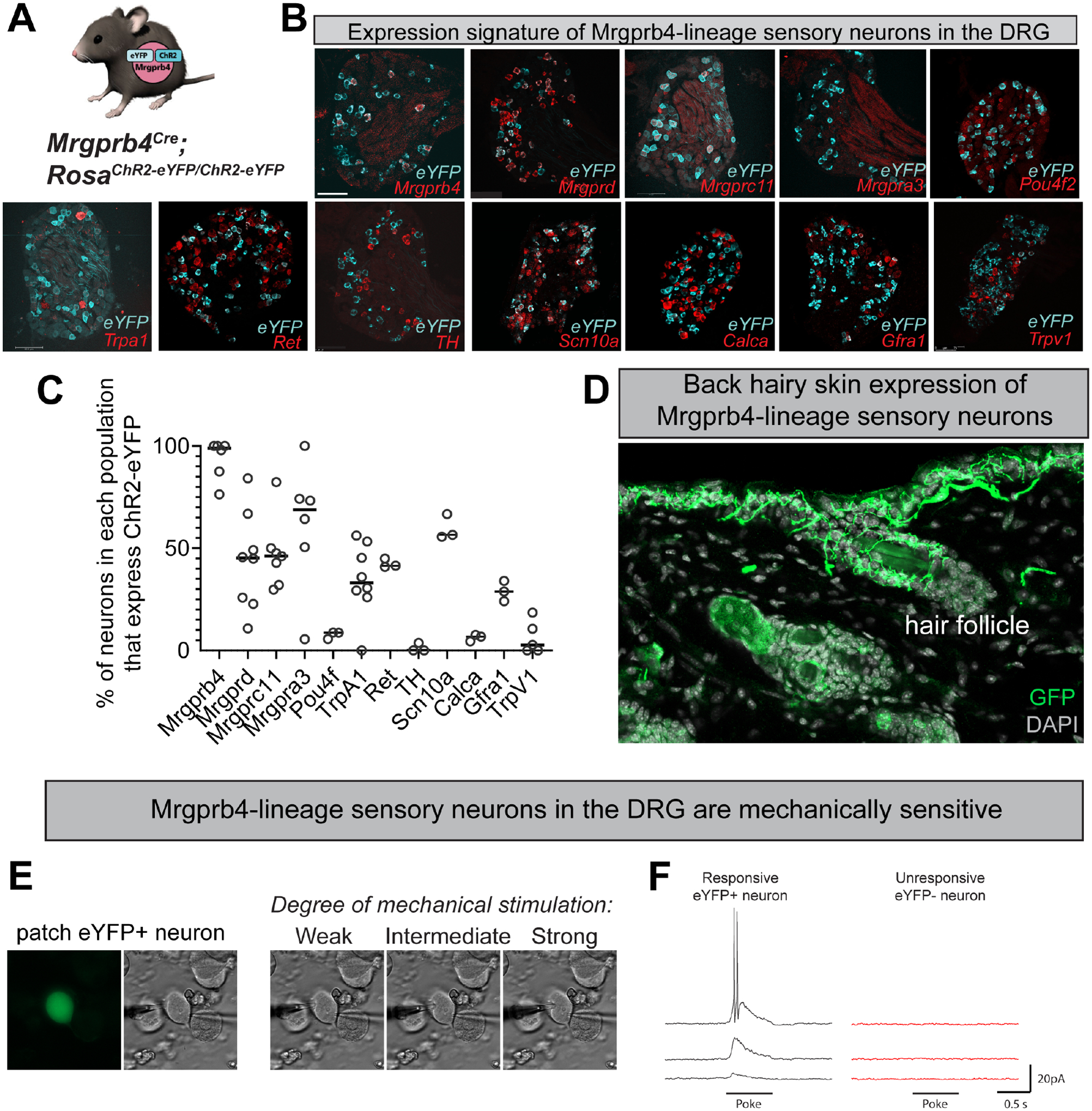
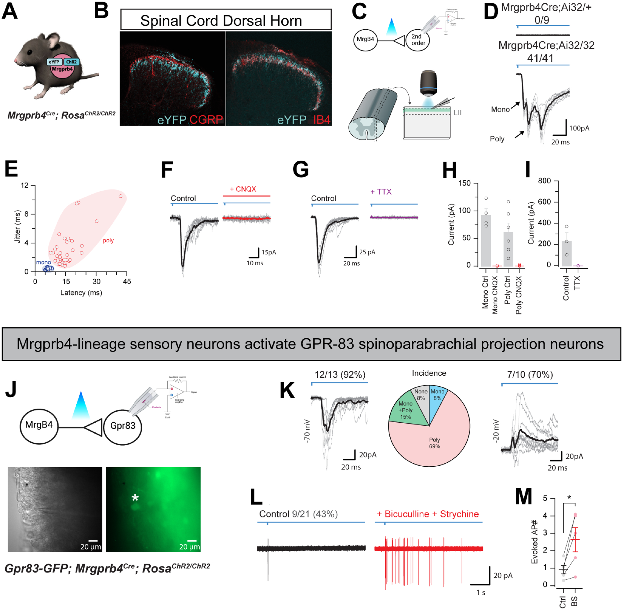
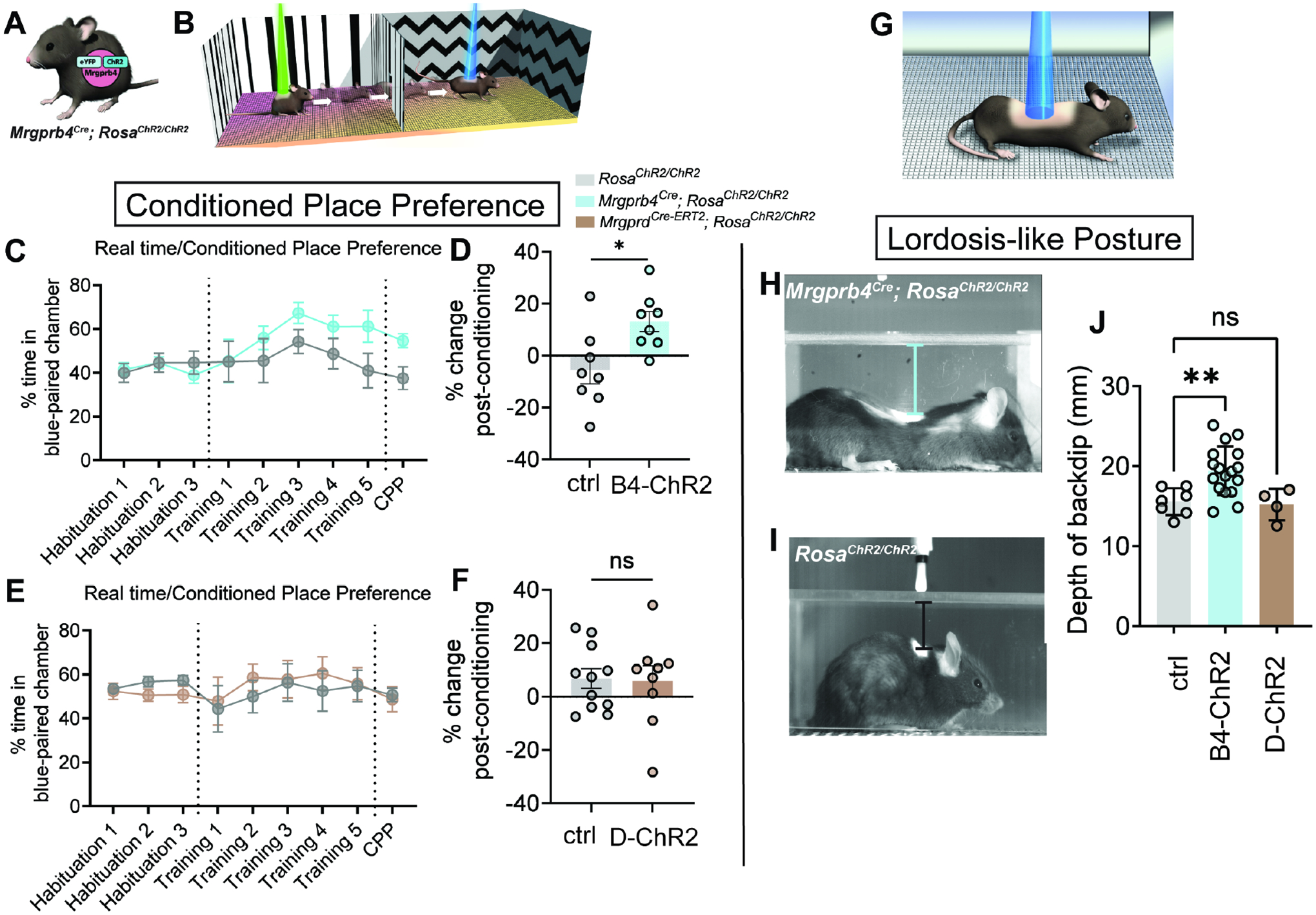
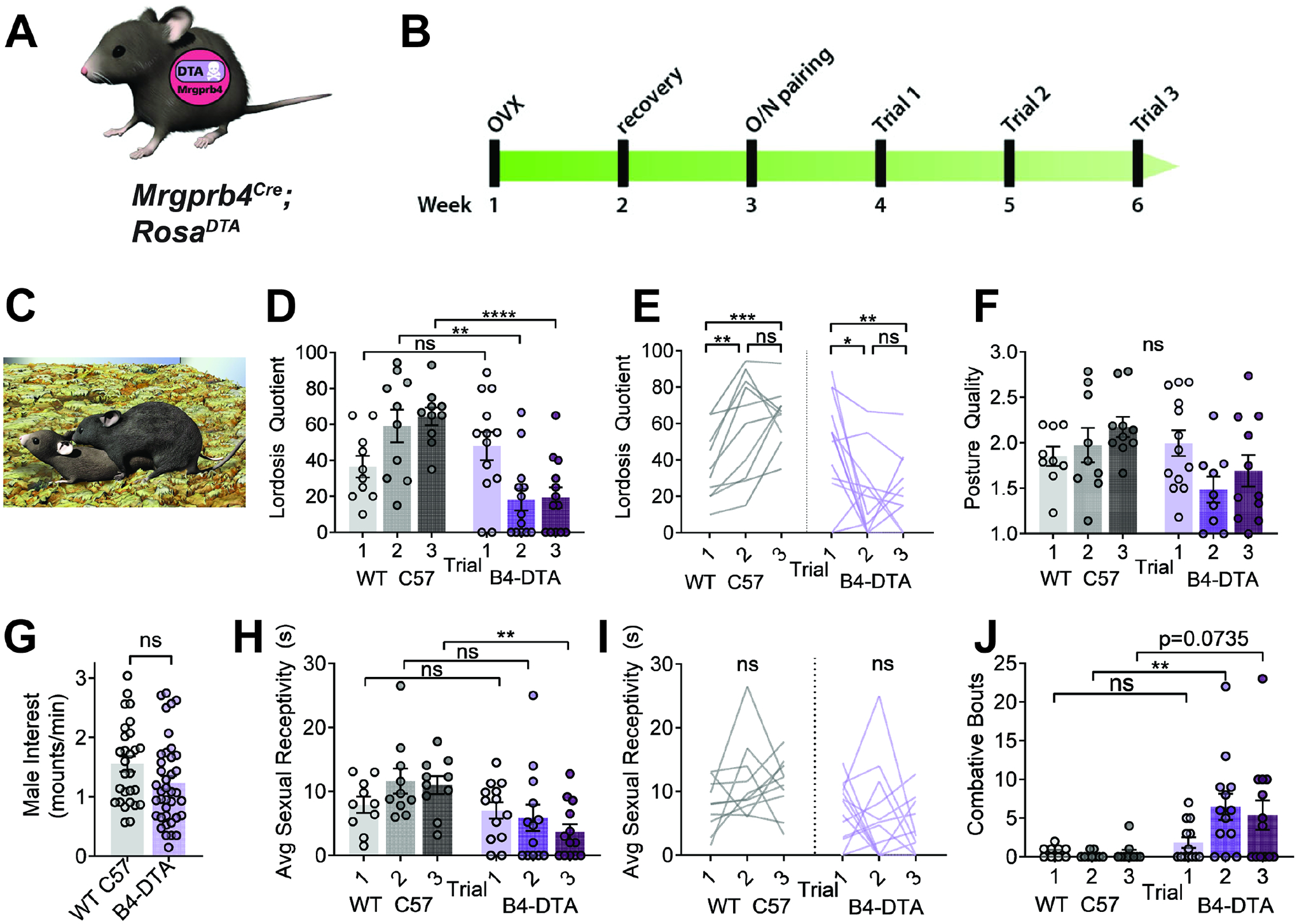
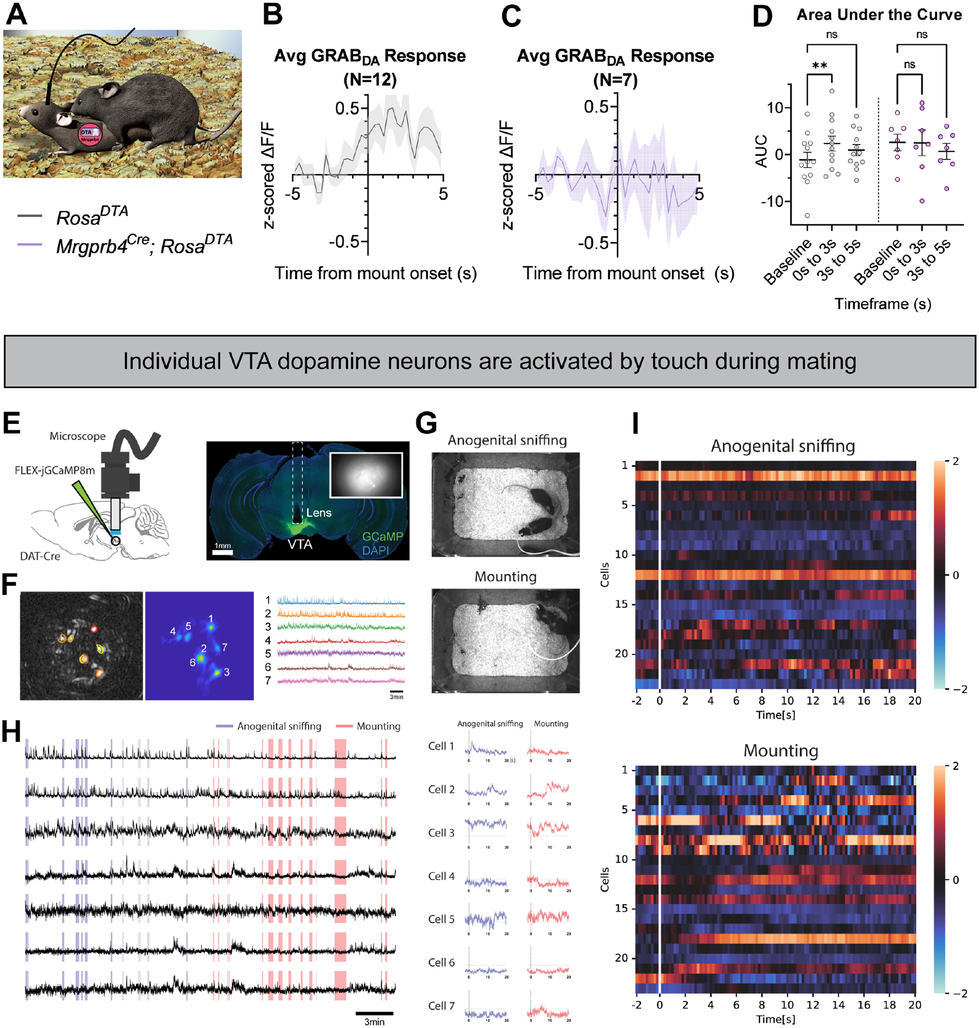
Pleasurable touch is paramount during social behavior, including sexual encounters. However, the identity and precise role of sensory neurons that transduce sexual touch remain unknown. A population of sensory neurons labeled by developmental expression of the G protein-coupled receptor Mrgprb4 detects mechanical stimulation in mice. Here, we study the social relevance of Mrgprb4-lineage neurons and reveal that these neurons are required for sexual receptivity and sufficient to induce dopamine release in the brain. Even in social isolation, optogenetic stimulation of Mrgprb4-lineage neurons through the back skin is sufficient to induce a conditioned place preference and a striking dorsiflexion resembling the lordotic copulatory posture. In the absence of Mrgprb4-lineage neurons, female mice no longer find male mounts rewarding: sexual receptivity is supplanted by aggression and a coincident decline in dopamine release in the nucleus accumbens. Together, these findings establish that Mrgprb4-lineage neurons initiate a skin-to-brain circuit encoding the rewarding quality of social touch.

Keywords: peripheral optogenetics; pleasure; reward pathway; spinal cord; touch.


Conflict of interest statement

Declaration of interests The authors declare no competing interests.

Figures

Figure 1:
Figure 1:. Genetic targeting strategy to optogenetically manipulate Mrgprb4-lineage touch neurons.
A) Mrgprb4Cre mice express ChR2-eYFP in a Cre-dependent manner. B) RNAscope in situ hybridization in DRG to quantify the expression of ChR2 in Mrgprb4+, Mrgprd+, Mrgprc11+, Mrgpra3+, Pou4f2+, Trpa1+, Ret+, TH+, Scn10a+, Calca+, Gfra1+, and Trpv1+ cells. Scale bars represent 100 μm. C) Percent expression of eYFP in different populations of DRG neurons from B. D) Expression of ChR2-eYFP in Mrgprb4-lineage nerve terminal endings in the hairy skin of the mouse back, labeled via immunofluorescence. E-F) Whole cell patch clamp recordings from individual eYFP+ Mrgprb4-lineage neurons demonstrating mechanical sensitivity. Increase in current response with increased mechanical stimulation.
Figure 2:
Figure 2:. Mrgprb4-lineage neurons activate downstream spinal circuits including Gpr83+ spinoparabrachial projection neurons.
A) Mrgprb4Cre mice express ChR2-eYFP in a Cre-dependent manner. B) Immunofluorescence staining in spinal cord dorsal horn. Mrgprb4+ terminals innervate lamina II, inferior to CGRP+ terminals in lamina I and overlapping with IB4+ terminals in lamina II. Scale bars represent 100 μm. C) Schematic illustrating spinal cord slice electrophysiological recordings from lamina II during optogenetic stimulation of terminals. D) Mono- or poly-synaptic light induced currents only in Mrgprb4Cre; RosaChR2/ChR2 mice. E) Quantification of light induced currents from Mrgprb4Cre; RosaChR2/ChR2 mice with jitter used to determine mono- or poly-synaptic transmission. F) Light-induced currents in Mrgprb4Cre; RosaChR2/ChR2 mice are abolished with CNQX. G) Light-induced currents are abolished in Mrgprb4Cre; RosaChR2/ChR2 mice with TTX. H,I) Quantification of data presented in F,G. J) Schematic, brightfield, and fluorescent images illustrating recording from GFP+ GPR83+ neurons during optogenetic stimulation of Mrgprb4-lineage neurons. K-M) Characterization of light-induced inputs to Gpr83+ putative spinoparabrachial projection neurons. K) Left: Whole-cell voltage clamp recording at −70 mV demonstrating excitatory inputs, Middle: Incidence of a monosynaptic only, polysynaptic only, or mono + polysynaptic excitatory input, Right: Whole-cell voltage clamp recording at −20 mV demonstrating inhibitory inputs. L) Cell-attached recording showing light-evoked action potential discharge under normal conditions (left) and following addition of bicuculline and strychnine (right). M) Quantification of data presented in L.
Figure 3:
Figure 3:. Focalized activation of Mrgprb4-lineage neurons in the back skin is rewarding and induces a lordosis-like posture in female mice.
A) Mrgprb4Cre; RosaChR2/ChR2 females and Cre-negative littermates are used in this assay B) Schematic illustrating our real time/conditioned place preference assay. C) Mrgprb4Cre; RosaChR2/ChR2 females gradually spend more time in the blue laser chamber compared to green laser chamber during the training days (lasers on) and D) exhibit a significantly greater change in time spent in the chamber they learned to associate with blue light on the test day (lasers off) compared to Cre-negative littermate controls. (*p=0.0139, unpaired t-test.) E,F) MrgprdCRE-ERT2; RosaChR2/ChR2 females do not develop a preference for the blue laser-paired chamber. H,I) Stills from high speed videography to closely examine the behavior during the preferable transdermal optogenetic activation of Mrgprb4-lineage neurons. Mrgprb4Cre; RosaChR2/ChR2 females (H) exhibit a striking lordosis-like dorsiflexion in response to Mrgprb4-lineage neuron activation, a behavior absent from (I) RosaChR2/ChR2 female littermates and MrgprdCRE-ERT2; RosaChR2/ChR2 females. J) This posture is quantified as the back’s maximum distance from the chamber ceiling over the course of 20s optogenetic stimulation (**p=0.0065, one-way ANOVA). All optogenetic stimuli are 35mW, pulsed at 10Hz sin wave.
Figure 4:
Figure 4:. Mrgprb4-lineage neurons are required for female sexual receptivity.
A) Graphic representing Mrgprb4Cre; RosaDTA mouseline, expressing DTA in a Cre-dependent manner to ablate Mrgprb4-lineage neurons. B) Timeline for assessing sexual receptivity with lordosis quotient (LQ) assay: all females are given two weeks to recover after ovariectomy (OVX), at which point estradiol and progesterone are administered to put them in behavioral estrus for an overnight pairing with a male. Hormones replaced prior to each LQ trial. C) Assays conducted in the male home cage in the dark cycle. Graphic depicts a female in sexually receptive lordosis posture in response to male mount D,E) Lordosis quotient scores for 3 sequential trials for WT C57 and Mrgprb4Cre; RosaDTA females. (Two-Way Repeated Measures ANOVA) D) On trials 2 and 3, Mrgprb4Cre; RosaDTA females exhibited significantly reduced sexual receptivity compared to WT females at the same trials (Trial 2 **p=0.0052; Trial 3 ****p<0.0001, Sidak’s multiple comparison test) E) Changes in individual mice across the three trials. WT females exhibit significant increase in sexual receptivity from trial 1 to trial 2 (**p=0.0055) and from trial 1 to trial 3 (***p=0.0009), whereas Mrgprb4Cre; RosaDTA females exhibit significant decrease in sexual receptivity from trial 1 to trial 2 (*p=0.0237) and from trial 1 to trial 3 (**p=0.0059), Tukey’s multiple comparisons test). F) Posture quality assessed on a scale from 1–3, where 1 is the minimum receptive posture and 3 is the most robust posture (details in methods) (ns, Two-Way Repeated Measures Mixed-effects analysis). G) Male mounting frequency was no different between Mrgprb4Cre; RosaDTA females and controls (ns, independent samples t-test). H) Average sexual receptivity, or duration maintained a receptive posture. (**p=0.0021, Two-Way Repeated Measures ANOVA, Sidak’s multiple comparisons test). I) Changes in sexual receptivity in individual mice across trials (ns, Tukey’s multiple comparisons test). J) Total number of combative bouts observed for each female in each trial. (**p=0.0096, Two-Way Repeated Measures Mixed-effects analysis, Sidak’s multiple comparisons test)
Figure 5:
Figure 5:. Activation of Mrgprb4-lineage neurons triggers dopamine release in the nucleus accumbens.
A) Schematic depicting the experimental setup. Female Mrgprb4Cre; RosaChR2/ChR2 mice with shaved backs, which had been injected with GRABDA to the NAc two weeks prior to testing, were placed under plastic chambers on a mesh platform. Pulsed blue (stimulating) or green (control) laser light (35mW, 10Hz sin wave) was shined to either the back skin (C-F) or skin surrounding the vagina (G-J) while recording GRABDA signals. B) Cannula placement and GRABDA expression in NAc C) ChR2-eYFP expression in Mrgprb4-lineage terminals in the back skin. D) Average GRABDA signal as z-score upon blue light (D) or green light (E) stimulation to the back. F) Average deltaF/F signals during pre-stimulation baseline (−5–0s) and during stimulation (0–6s) for each animal. Repeated measures two-way ANOVA revealed significant interaction between wavelength and time (p=0.0161) and Sidak’s multiple comparison test revealed significant difference in blue light (**p=0.0014) but not green light (p=0.9411) pre-stim compared to during stimulation. G-J) Average GRABDA signal as z-score upon blue light (H) or green light (I) stimulation to the anogenital region. J) Average deltaF/F signals during pre-stimulation baseline (−5–0s) and during stimulation (13–20s) for each animal. Repeated measures two-way ANOVA revealed significant interaction between wavelength and time (p=0.0110) and Sidak’s multiple comparison test revealed significant difference in blue light (***p=0.0003) but not green light (p=0.8547) pre-stim compared to during stimulation.
Figure 6:
Figure 6:. Mrgprb4-lineage neurons are required for dopamine release in the nucleus accumbens during sexual mounts.
A) Graphic depicting experimental setup. Female Mrgprb4Cre; RosaDTA mice or littermate RosaDTA controls, which had been injected with GRABDA to the NAc over two weeks prior to testing were paired with males for a mating assay. Females were ovariectomized and hormone primed to be in a state of behavioral estrus for testing. GRABDA signals were recorded for the entire pairing and analyzed surrounding male mounts. B) Average z-score traces surrounding mount onset (T=0) for littermate RosaDTA controls (gray) (N=12) and C) Mrgprb4Cre; RosaDTA females (purple) (N=7) D) Area under the curve for Z-scored signals at pre-mount baseline (−5 to −2s), 0–3s post-mount, and 3–5s post-mount for each animal. Repeated measures two-way ANOVA revealed a significant interaction between genotype and time (p=0.037), and Sidak’s multiple comparison test revealed significant difference in GRABDA signal between baseline and 0–3s post mount for control (**p=0.0024) but not Mrgprb4Cre; RosaDTA females (p=0.9945). E) Schematic of a miniaturized one-photon fluorescence microscope and calcium imaging. A DAT-Cre animal was unilaterally injected with jGCaMP8m virus into the left hemisphere of the ventral tegmental area (VTA; AP:−3.20 mm, ML: −0.40 mm, DV: −4.40 mm). DAPI-stained (blue) coronal section of DAT-Cre mouse showing GCaMP8m fluorescence (green) and the location of a 0.6mm-diameter gradient index (GRIN) lens implant (dashed lines). The inset image indicates the imaging field from the lens. F) A cell contour map (left) and CNMFe-processed image of the neurons located in the VTA (middle) with corresponding raw calcium traces (right). G) Sample video frames of anogenital sniffing and mounting. The animal poses were manually annotated on a frame-by-frame by two human annotators. H) Raw calcium traces from 7 dopamine cells in VTA (black lines) with two manual behavior annotations: anogenital sniffing (blue; 14 events) and mounting (red; 16 events). Median z-scored calcium signals of each dopamine cell at the time of anogenital sniffing (blue; left) or mounting (red; right). I) Representative calcium activity of VTA dopamine neurons during anogenital sniffing or mounting (n=23 cells, 3 mice).

References

    1. Orefice LL, Mosko JR, Morency DT, Wells MF, Tasnim A, Mozeika SM, Ye M, Chirila AM, Emanuel AJ, Rankin G, Fame RM, Lehtinen MK, Feng G, Ginty DD. Targeting Peripheral Somatosensory Neurons to Improve Tactile-Related Phenotypes in ASD Models. Cell. 2019;178(4):867–86 e24. Epub 2019/08/10. doi: 10.1016/j.cell.2019.07.024. - DOI - PMC - PubMed
    1. Orefice LL, Zimmerman AL, Chirila AM, Sleboda SJ, Head JP, Ginty DD. Peripheral Mechanosensory Neuron Dysfunction Underlies Tactile and Behavioral Deficits in Mouse Models of ASDs. Cell. 2016;166(2):299–313. Epub 2016/06/14. doi: 10.1016/j.cell.2016.05.033. - DOI - PMC - PubMed
    1. Peled-Avron L, Shamay-Tsoory SG. Don’t touch me! autistic traits modulate early and late ERP components during visual perception of social touch. Autism Res. 2017;10(6):1141–54. Epub 2017/03/25. doi: 10.1002/aur.1762. - DOI - PubMed
    1. Hart AS, Rutledge RB, Glimcher PW, Phillips PE. Phasic dopamine release in the rat nucleus accumbens symmetrically encodes a reward prediction error term. J Neurosci. 2014;34(3):698–704. Epub 2014/01/17. doi: 10.1523/JNEUROSCI.2489-13.2014. - DOI - PMC - PubMed
    1. Mohebi A, Pettibone JR, Hamid AA, Wong JT, Vinson LT, Patriarchi T, Tian L, Kennedy RT, Berke JD. Dissociable dopamine dynamics for learning and motivation. Nature. 2019;570(7759):65–70. Epub 2019/05/24. doi: 10.1038/s41586-019-1235-y. - DOI - PMC - PubMed

Publication types

LinkOut - more resources